
Figure 12.
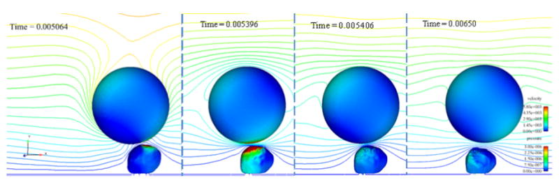
Four time steps of the collision simulation in which bonds formed between the cancer cell and white blood cell. Time steps shown are: a) before bonds form, b) when the number of bonds is a maximum, c) immediately after the bonds break, and d) one millisecond later. The cells are contoured by pressure (in kg/μm-s2) and a center clip plane is contoured by velocity magnitude (in μm/s). Flow direction is left to right.