
Figure 2. Identification of kaempferol metabolites from CaCo-2/TC7 cells and associated media.
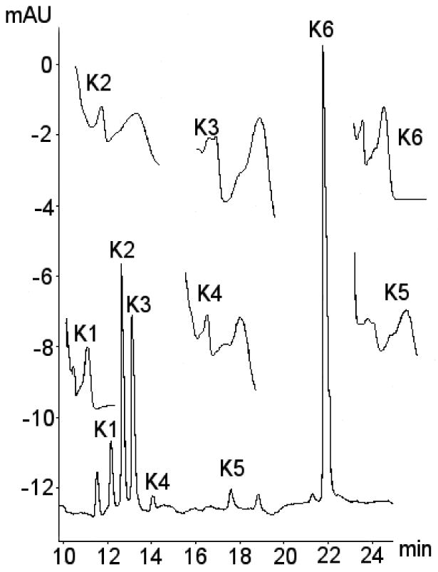
Typical chromatogram showing metabolites of kaempferol. The identities of metabolites were confirmed using a combination of retention time, selected ion monitoring LC/MS, tandem mass spec with metal complexation, kaempferol-3-glucuronide pure authentic standard, enzyme hydrolysis with sulphatase and glucuronidase and UV spectra. Suspected metabolites labelled K1-K5, with kaempferol aglycone labelled K6. UV spectra for the various metabolite peaks also shown with UV peak absorbance at around 250-280 nm and 350-400 nm.