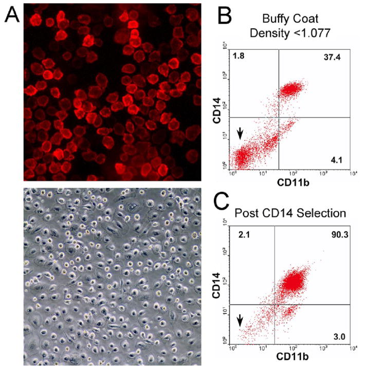
Figure 1. Isolation of osteoclast precursors from human peripheral blood.
A. Cells isolated by anti-CD14 immuno-magnetic separation from the mononuclear fraction of human peripheral blood. Fluorescence micrograph (top frame) of CD14-selected cells labeled with Cy5-conjugated anti-CD14 antibody (field 220 μm square) four hours after isolation, and a phase micrograph (lower frame) of 6 day culture (field 880 μm square). In each case, non-adherent cells were washed from the cultures after plating.
B,C. Expression of CD14 and CD11b by human peripheral blood mononuclear cells and cells isolated by immuno-magnetic separation. Mononuclear cells (B), isolated by density gradient centrifugation from human buffy coat cells, and cells (C) from the same buffy coat after CD14 immuno-magnetic selection are analyzed by flow cytometry as described in Methods. Compared to the results for unselected peripheral blood mononuclear cells (B), after immuno-magnetic selection (C), the fraction of cells not labeled by either CD14 or CD11b is greatly reduced, as expected (arrows). After selection, over 95% of cells labeled with CD14 or CD11b, while lymphocyte markers were present on <2% of cells (see text).

