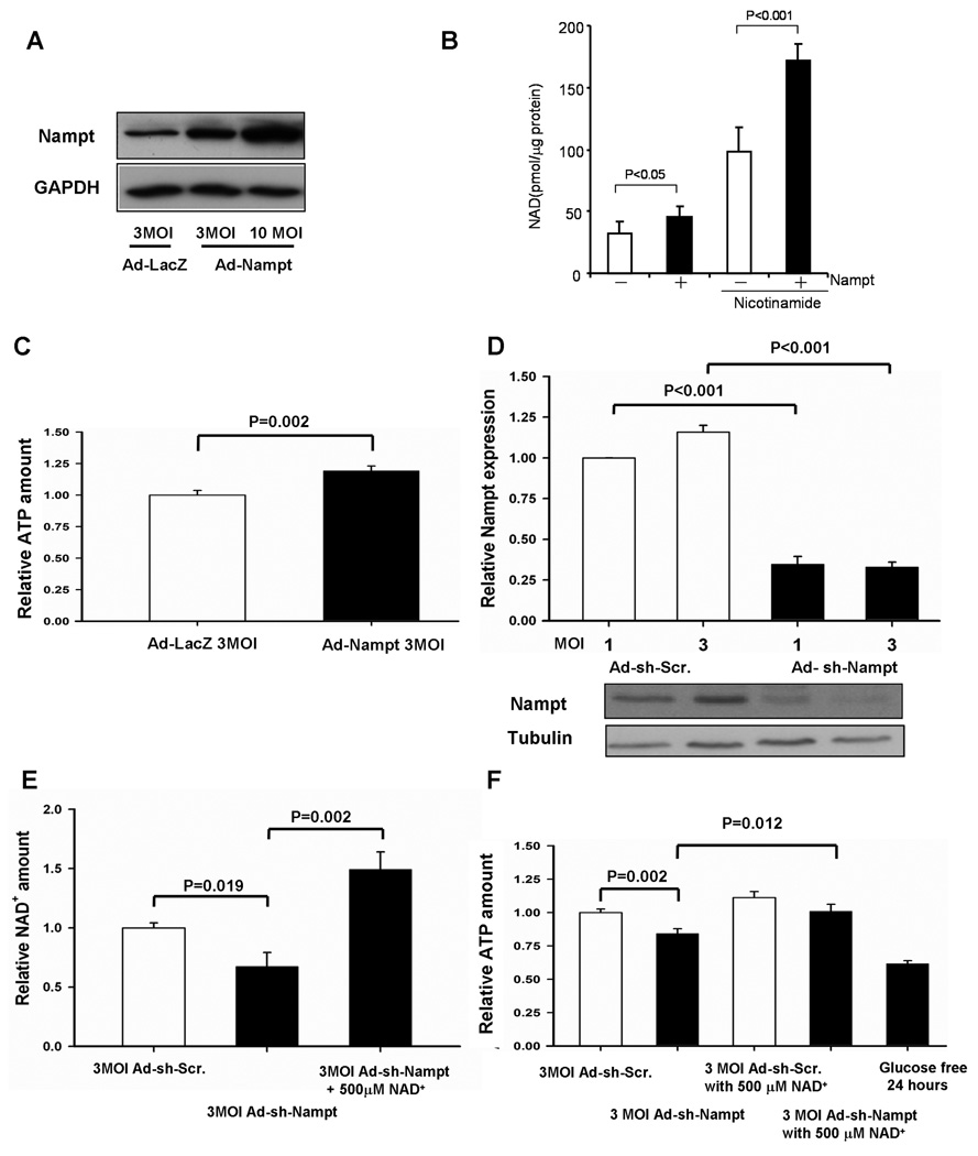
Figure 2. Nampt regulates NAD+ and ATP levels in cardiac myocytes.
Cardiac myocytes were transduced with Ad-LacZ, Ad-shRNA-scramble, Ad-Nampt or Ad-shRNA-Nampt at indicated MOIs. In A and D, cell lysates were subjected to immunoblot analyses with anti-Nampt, anti-GAPDH and anti-tubulin antibodies. In B, C, E and F, Cellular [NAD+] and [ATP] were measured using the EnzyChromTM NAD+/NADH Assay Kit and ATP Bioluminescent Assay Kit, respectively. In B, cells were cultured with either normal culture media or those supplemented with 20 mM nicotinamide for 16 hours. In C-F, the level of Nampt, [NAD+] and [ATP] in control virus transduced myocytes is expressed as 1. In E and F, cardiac myocytes were cultured with or without 500 µM NAD+ or glucose free medium for 24 hours. Data represent the mean of four experiments ± SEM.

