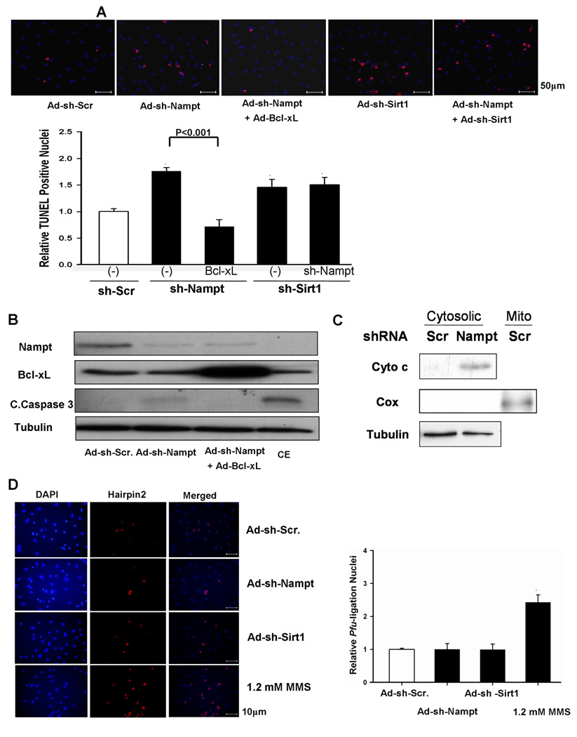
Figure 3. Knockdown of Nampt causes apoptotic cell death in cardiac myocytes.
Cardiac myocytes were transduced with Ad-shRNA-scramble, Ad-shRNA-Nampt, or Ad-shRNA-Sirt1 alone or in combination. In some cases, Ad-LacZ or Ad-Bcl-xL was co-transduced. Myocytes were then cultured for 96 hours. (A) (upper) TUNEL staining of myocytes. Nuclei were counterstained with DAPI. (lower) Relative TUNEL-positive nuclei among different groups. The value in control myocytes was designated as 1. (B) Cell lysates were subjected to immunoblot analyses with anti-Nampt, anti-Bcl-xL, anti-cleaved caspase-3 (C. Caspase-3), and anti-tubulin antibodies. Myocytes treated with chelerythrine (CE, 10 µM for 2 hours) were used as a positive control for apoptosis. (C) Cardiac myocytes were transduced with Ad-shRNA-scramble (Scr) or Ad-snRNA-Nampt (Nampt) (3 MOI) and cultured for 5 days. The cytosolic fraction was subjected to immunoblot analyses with anti-cytochrome c (Cyto c), anti-cytochrome c oxidase I (Cox) and anti-tubulin antibodies. The mitochondrial fraction (Mito) was included as a positive control for Cox. (D) Nuclei were stained using the Texas Red labeled blunt end probe synthesized with pfu. Myocytes treated with 1.2 mM methylmethane sulfonate (MMS) for three hours were used as a positive control for necrotic cell death. (left) Representative images of DAPI and hairpin 2 staining and merged images. (right) Quantitative analyses of hairpin 2 positive cardiac myocytes. The value in control myocytes was designated as 1. * p<0.05 vs Ad-shRNA-scramble. The results are mean of 4 experiments.

