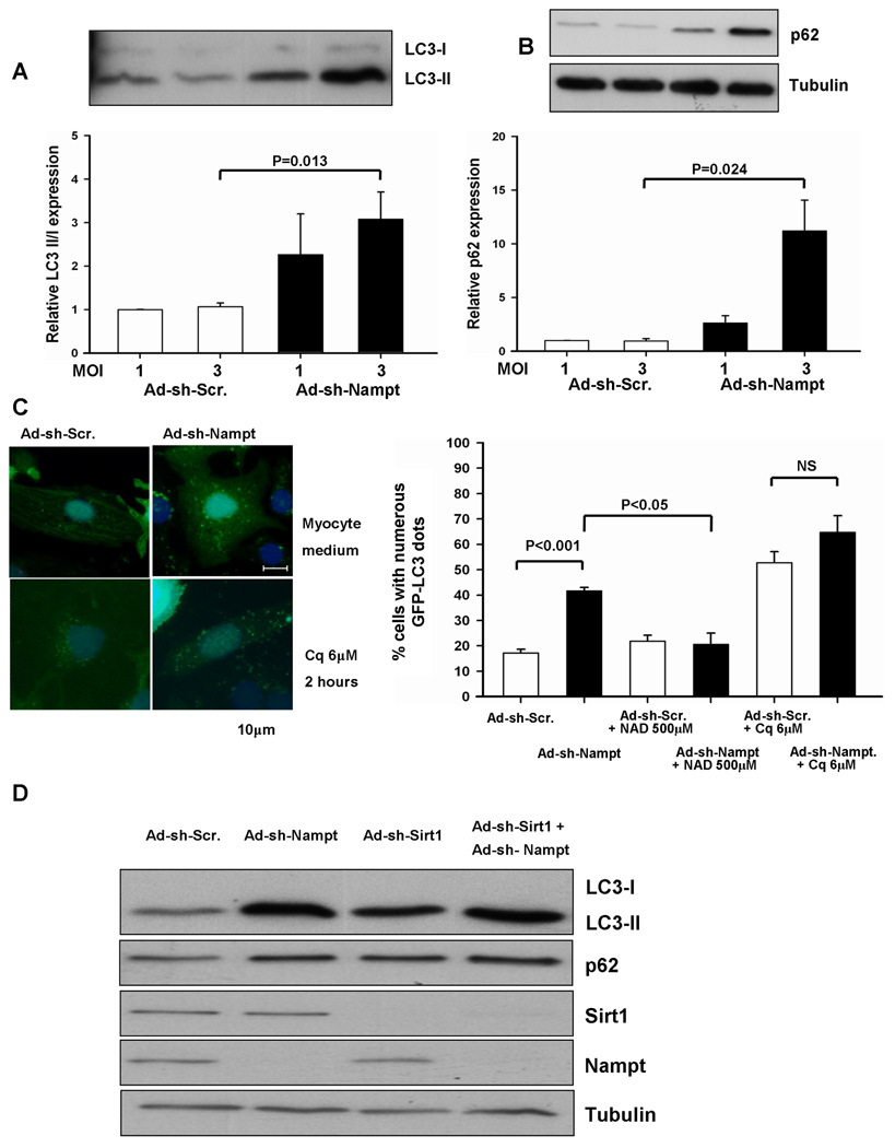
Figure 4. Knockdown of Nampt affects autophagic flux, in a manner similar to chloroquine treatment.
(A and B) Myocytes were transduced with Ad-shRNA-scramble (Ad-sh-Scr.) or Ad-shRNA-Nampt (Ad-sh-Nampt) at indicated MOIs and cultured for 96 hours. Cell lysates were subjected to immunoblot analyses with anti-LC3 (A), anti-p62 (B), and anti-tubulin (B) antibodies. The values of LC3-II/LC3-I and p62 expression in myocytes transduced with Ad-shRNA-scramble are designated as 1. Experiments were conducted 3 times. (C) Myocytes were transduced with Ad-GFP-LC3 (10 MOI) and either Ad-shRNA-scramble or Ad-shRNA-Nampt at 3 MOI and cultured for 96 hours. Some myocytes were treated with chloroquine (Cq, 6 µM) for 2 hours or NAD+ (500 µM) for 24 hours. (left) Representative images of GFP-LC3 staining are shown. (right) The percentage of cells with punctate GFP-LC3 staining is shown. N=4. (D) Myocytes were transduced with Ad-shRNA-scramble, Ad-shRNA-Nampt, or Ad-shRNA-Sirt1 alone or in combination and cultured for 96 hours. Cell lysates were subjected to immunoblot analyses with anti-LC3, anti-p62, anti-Sirt1, anti-Nampt, and anti-tubulin antibodies. The results shown are representative of three experiments.

