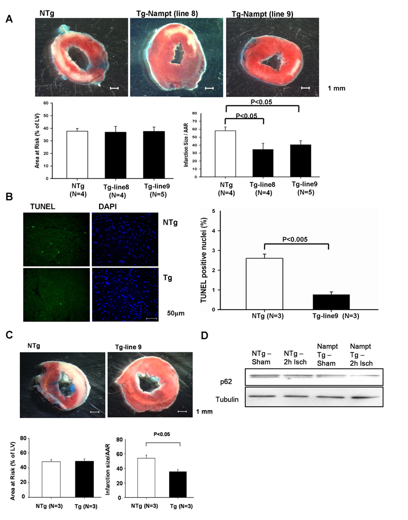
Figure 6. I/R injury is attenuated in Tg-Nampt mice.
(A) Tg-Nampt and NTg mice were subjected to 45 minutes of ischemia and 24 hours of reperfusion. (upper) Gross appearance of LV myocardial sections after Alcian blue and triphenyltetrazolium chloride (TTC) staining. (lower left) The area at risk (AAR) (% of LV) was comparable between NTg and Tg-Nampt. (lower right) The infarction area/AAR was significantly smaller in Tg-Nampt than in NTg mice. (B) (left) LV myocardial sections were subjected to TUNEL and DAPI staining. Representative images of the staining in the border zone are shown. (right) The number of TUNEL-positive myocytes is expressed as a percentage of total nuclei detected by DAPI staining. (C and D) Tg-Nampt and NTg mice were subjected to 2 hours of ischemia. (C) (upper) Gross appearance of LV myocardial sections after Alcian blue and TTC staining. (lower left) The area at risk (AAR) (% of LV) was comparable between NTg and Tg-Nampt. (lower right) The infarction area/AAR was significantly smaller in Tg-Nampt than in NTg. (D) Expression of p62 and tubulin at the baseline and after 2 hours of ischemia was determined by immunoblot analyses. The results shown are representative of five experiments.

