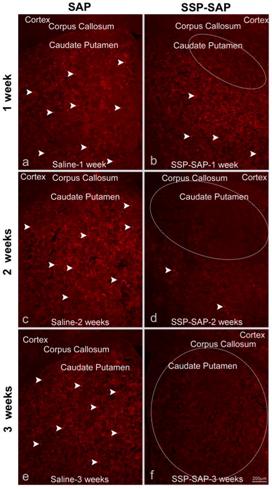
Figure 2.

Ablation of striatal neurokinin-1 receptor-expressing neurons using SSP-SAP. Animals were intrastriatally injected with SSP-SAP [4ng, 1 ul volume] (b, d and f) or saporin (SAP) [4ng, 1 ul volume] ) on the contralateral side (a, c and e). Animals were sacrificed at 1, 2 or 3 weeks after the intrastriatal injections and sections of striatal tissue were immunostained with an antibody against the neurokinin-1 receptor. Microinjection of SAP alone had no effect on neurokinin-1 receptor immunostaining in the striatum. Note the lack of immunostaining for the neurokinin-1 receptor at 3 weeks after the injection with SSP-SAP (f). White dotted circle indicates the region lacking neurons that express the neurokinin-1 receptors. White arrows indicate immunostaining of representative neurokinin-1 receptor-bearing interneurons. Magnification bar is 200 μm.
