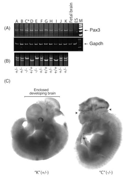
FIG. 4.
Exencephalic embryos were detected as early as E11.5, although Pax3 expression was not altered by the absence of TEAD2. (A) Total RNA was prepared from 11 embryos at E11.5 that were obtained from a single heterozygous female mated to a heterozygous male. This RNA was assayed for Pax3 sequences using RT-PCR. RNA from E13.5 fetal brain served as a positive control, while RNA from ES cells served as a negative control. One embryo that exhibited exencephaly (asterisk) is shown in panel C. (B) Multiplex PCR analysis was used to determine the genotype of each embryo. (C) One normal (Tead2+/−) and one exencephalic (Tead2−/−) embryo from the same litter at E11.5 are shown using bright-field illumination. Arrows indicate sites where neural tube failed to close.

