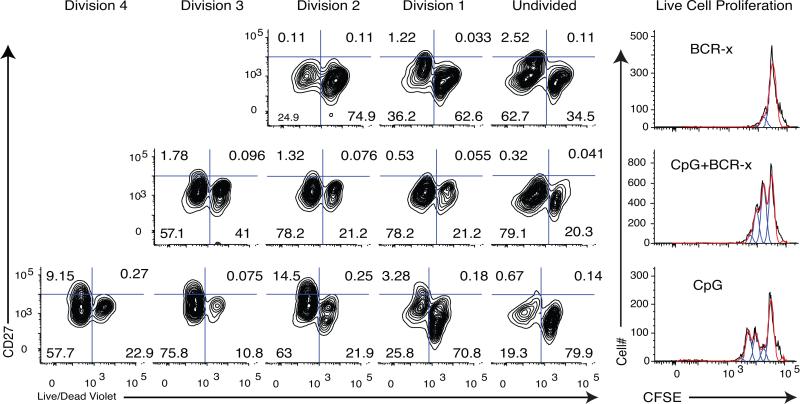
Figure 7. Increased cell death in later divisions of CpG+CK+BCR-x stimulated cells.
IgMneg memory B cells were isolated from the donor and split into treatment groups, with medium alone (Unstimulated), BCR-x, CpG+CK, and CpG+CK+BCR-x. After 72 hours in culture, the cells were stained for cell surface CD27 and live/dead violet dye (representative of 3 experiments). Dead cells retain this dye, appearing to the right in these figures. CpG+CK treatment produced more dead cells in Undivided and Division 1 populations. Addition of BCR-x to CpG+CK changed this profile to favor more live cells in undivided populations and more dead cells in later divisions. This suggests that reduced numbers of high-rate IgG-SC and reduced B cell blasts in CpG+CK+BCR-x may be due to higher death rates.

