Figure 1.
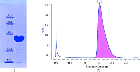
(a) 12% SDS–PAGE of pure recombinant MpgS from T. thermophilus HB27. Lane 1, low-range molecular-weight markers (Bio-Rad): from the highest to the lowest weight, phosphorylase b (97.4 kDa), bovine serum albumin (66.2 kDa), ovalbumin (45 kDa), carbonic anhydrase (31 kDa), soybean trypsin inhibitor (21.5 kDa) and lysozyme (14.4 kDa). Lane 2, MpgS monomer migrating according to its molecular weight (43.5 kDa). (b) Elution profile of MpgS loaded onto an analytical 2.4 ml Superdex 200 3.2/30 PC column. The elution volume suggests that the protein is most likely to be in the dimeric state. mAU stands for milliunits of absorption.

