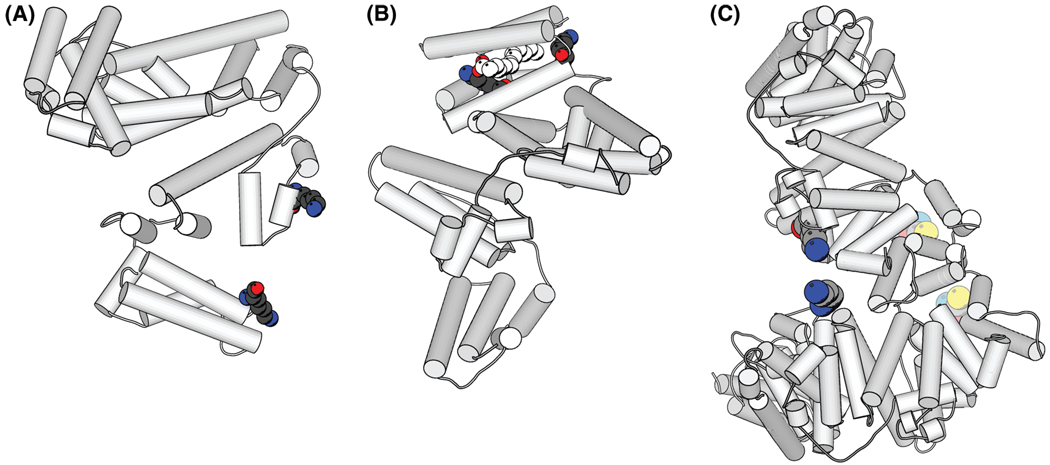
Figure 5.
Crystal structure of HSA (PDB code 1n5u). The location of the cross-linked peptides are highlighted in color in the ribbon and space fill diagram. (a) Intramolecular cross-linking on the surface of HSA. Balls represent the cross-linked peptide FK̃ (LKE)DLGE. (b) The intramolecular cross-linked peptides K̃ (HKPK)QTALVE within HSA. The white balls represent overlap with the ligand (myristic acid) observed in the crystal structure. The cross-linker SAMS apparently occupies the space of the ligand. (c) The cross-linked HSA dimer. Bright colored balls represent the cross-linked amino group in lysine in RYK̃ (SKDVCK)AAFTE. Pale colored balls represent the cross-linked thiol in Cys (34).26 This is a hypothetical structure of the HSA dimer (c) to illustrate the feasibility of the links. The plots were generated with Molscript.24 Atoms in the structure: C, light gray; O, red; N, sky blue; S, yellow.

