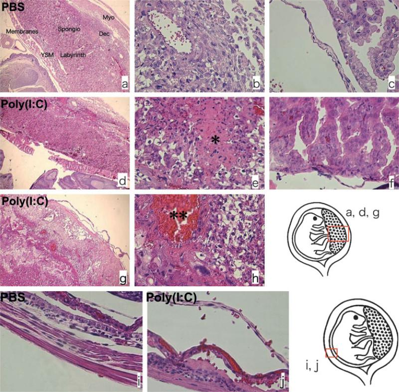
Fig. 1.
Poly[I:C] injection induced morphologic change at feto-maternal interface; 4.5 mg/kg Poly[I:C] or PBS was injected i.p. to wild type mice on 16.5 dpc and killed after 4 hr. Hematoxylin and eosin staining was performed for histopathologic analysis. The feto-maternal interface from PBS-treated animals kept its morphologic integrity (a–c and i). On the contrary, the feto-maternal interface from Poly[I:C] treated mice lost the integrity (d–h and j). Necrosis (e*), inflammation (e–g), hemorrhage (h**) and edema (f and j) were apparent in the decidua, the yolk sac membrane, chorion and amniotic membrane of poly(I:C) treated animals. The inflammation is accompanied by polymorphonuclear leukocytes infiltration (e–g). V: blood vessel, am: amniotic membrane, ysm; yolk sac membrane, ch: chorion, dec: decidua, myo: myometrium. Original magnification; a, d, g: 40×, b, c, e, f, h, i, j: 400×. Data are representative of at least three mice from same treatments.

