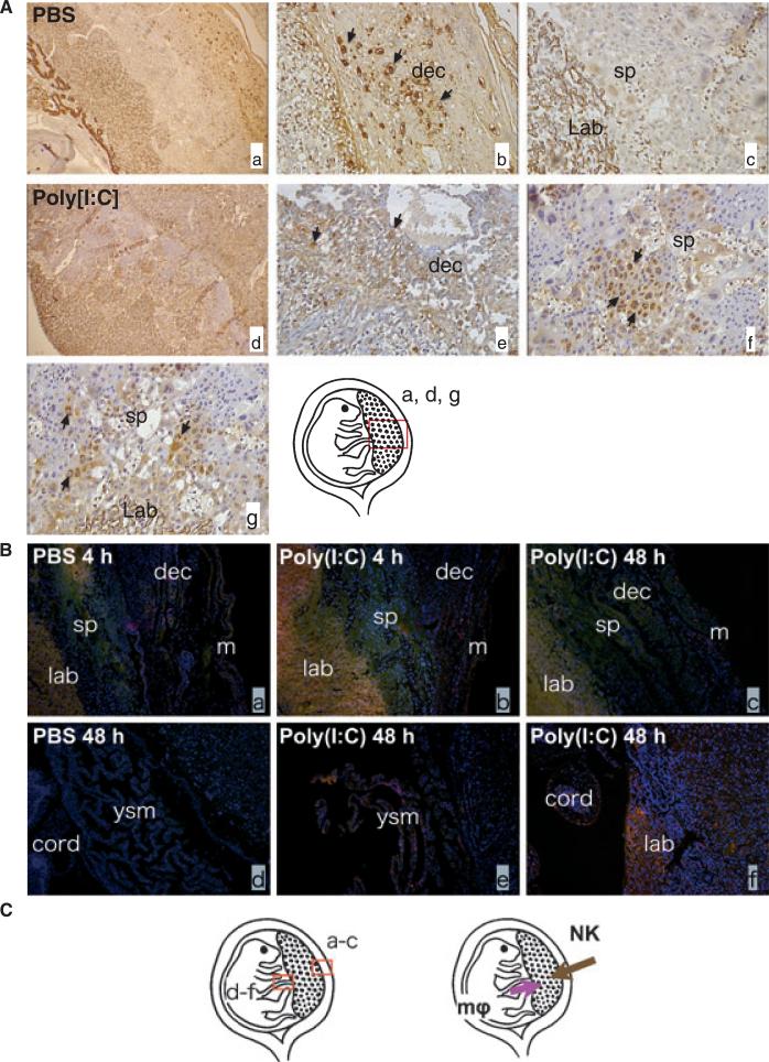
Fig. 2.
Poly[I:C] injection induced NK cells and macrophages infiltration into the placenta; 4.5 mg/kg Poly[I:C] or PBS was injected i.p. to wild type mice on 16.5 dpc. Mice were killed after either 4 or 48 hr. Utero-placental units were collected. Immunohistochemical analysis of the feto-maternal interface using lectin (for uNK cells; brown) or anti f4/80 antibody (for Macrophages; pink) were performed. (A) In PBS-treated mice, uterine NK cells were restricted to the decidua (Ab) and no NK cells were seen in spongiotrophoblast and labyrinth, (a–c). The distribution of NK cells changed in Poly[I:C] treated mice (d). Fewer NK cells were found in decidua (e) and more cells were found in spongiotrophoblast (f) and labyrinth zone (g). Slides were counterstained with hematoxylin (purple). (B) Macrophages were identified mainly in the outer myometrium and rarely in the decidua and were never observed in spongiotrophoblast and labyrinth in PBS-treated control animals (a). Macrophage infiltration from the maternal side toward the placental zone was not observed up to 48 hr after Poly[I:C] injection (b and c). Instead, when we observed the fetal side of the placenta at 48 hr post injection, we found macrophages infiltrated from the fetal side (umbilical cord and yolk sac membrane) toward the placenta zone (e and f). No such infiltration was found in PBS-treated animals (d). Slides were counterstained with Hoechst (blue nuclei). ysm, yolk sac membrane; m, myometrium; dec, decidua; sp, spongio; trophoblast, lab, labyrinth; cord, umbilical cord. (C) Schematic localization for uterine NK cells and macrophages and the direction of their infiltrations toward placental zone. Original magnification; 2A a and d: 40X, 2Ab, c, e, f and g: 2B a–f; 400X. Data are representative of at least three mice from same treatments.

