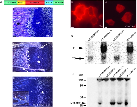
FIG. 1.
Transgene construction and expression of MT1-MMP in skeletal tissues. (A) Linear diagram of the collagen II/MT1-MMP transgene used for expression of MT1-MMP in cartilage tissue. Briefly, the mouse MT1-MMP cDNA (red) was placed under control of the type II collagen promoter/enhancer (green). Also shown is the β-globin intron (yellow) and the SV40 poly A signal sequence (blue). (B) Rat chondrosarcoma cells transfected with the transgene construct show abundant membrane-associated immunoreactivity when reacted with MT1-MMP–specific antibodies. Compare with C, depicting cells transfected with empty vector. (D) Northern blot of total RNA isolated from neonate mice reacted with an MT1-MMP–specific probe detects the endogenous transcript “E” and the transgene derived transcript “TG.” Lane 1, MT1-MMP−/−;T+ mouse; lane 2, MT1-MMP+/−;T+ littermate; lane 3, MT1-MMP−/−;T+ littermate; lane 4, MT1-MMP+/+ littermate. (E) H&E-stained femur from neonate control mouse showing the primary ossification center (asterisk) and the prospective epiphyseal growth plates “GP” of the distal condyle. (F) Dark field in situ hybridization image from the same block reacted with an mt1-mmp–specific antisense probe. Note the signal in periosteum (arrows) and the primary spongiosa (arrowheads). Signal is also detected in the proliferation zone of the prospective growth plate, although to a lesser extent than elsewhere (asterisk). (G) Section from femur of an MT1-MMP−/−;T+ mouse reacted with an antisense mt1-mmp probe. Note that expression is now restricted predominantly to the cartilage tissue (asterisks), whereas less, but still detectable, signal is found in the ossifying tissue (arrows inset in G, showing periosteal “P” and marrow signal between trabecular bone “TB”). (H) Immunoblot of costal chondrocyte extracts from neonate mice detecting MT1-MMP. Lane 1, MT1-MMP−/−;T+ mouse; lane 2, MT1-MMP−/− mouse; lane 3, MT1-MM+/−;T+ littermate; lane 4, MT1-MMP+/− littermate; lane 5, MT1-MMP+/+ littermate. Scale bars: (B and C) 10 μm; (E–G) 100 μm.

