FIG. 6.
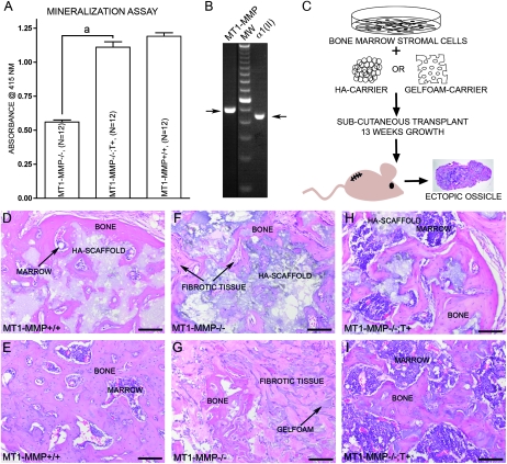
Expression of type II collagen in BMSCs and analysis of their osteogenic potential. (A) BMSCs derived from either MT1-MMP−/−, MT1-MMP−/−;T+, or MT1-MMP+/− mice show an increasing ability to form in vitro mineralized matrix in osteoconductive medium when measured as alizarin red retention. (B) In accordance with the results observed by in situ hybridization, immunostaining, and in the mineralization assay, BMSCs from wildtype mice show MT1-MMP and type II collagen–specific mRNA by RT-PCR (samples run on the same gel, but not adjacent lanes). This observation explains the expression derived from the collagen II–MT1-MMP transgene and the resulting MT1-MMP expression in BMSCs isolated from MT1-MMP−/−;T+ mice. (C) To test whether the osteogenic potential observed in vitro is biologically relevant, BMSCs on osteoconductive media were implanted in nude mice and allowed to form ectopic ossicles. (D and E) Cells derived from wildtype mice showed the ability to form both bone and marrow in hydroxyapatite (D) or Gelfoam scaffolds (E). Cells derived from MT1-MMP−/− mice failed almost completely to form bone in either hydroxyapatite (F) or Gelfoam (G) osteoinductive carriers nor did they support formation of marrow. Instead, extensive fibrosis was observed (arrows) (F and G). When MT1-MMP−/− cells expressed the collagen II–driven MT1-MMP transgene, the osteogenic potential was restored, and the cells generated abundant bone and marrow with both hydroxyapatite (H) and Gelfoam (I) as scaffolds, thus showing that the type II collagen promoter is active in BMSCs. Scale bar: (D–I) 100 μm. aStatistical significance (p < 0.001).

