
Figure 2.
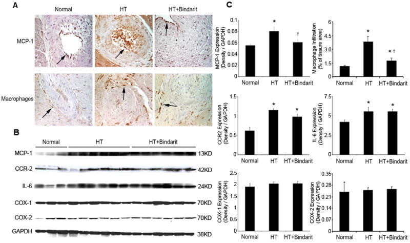
The expression of myocardial MCP-1, its receptor CCR2, cyclooxygenase (COX)-1, COX-2, IL-6, and macrophage infiltration in normal, hypertension (HTN), and HTN+bindarit pigs detected by immunohistochemistry. A. Representative images (magnification ×40) of MCP-1 and macrophage staining (brown, arrows), showing increase in HTN and decrease by bindarit. B. Western blots, showing increased expression of MCP-1, CCR-2 and IL-6 in both HTN and HTN+bindarit. COX-1 and COX-2 expression was similar among the groups. C. Quantitation and densitometry normalized by GAPDH. * p<0.05 vs. normal and † p<0.05 vs. HTN.