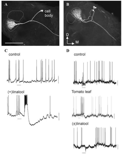
Figure 1.

Laser-scanning confocal microscopic images of female-specific output neurons with arborizations confined to the lateral large female glomerulus (latLFG, A) and the medial large female glomerulus (medLFG, B). Dotted lines indicate the outline of the female-specific glomeruli. Scale bar, 200 μm. D, dorsal; M, medial. C, D: Electrophysiological recordings obtained from neurons in a latLFG (C) and a medLFG (D) in response to the odors indicated at a concentration of 10-3 vol/vol. Neurons were stimulated as described previously.17
