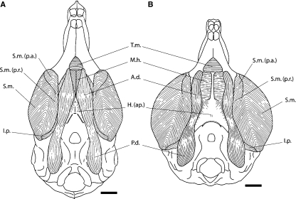
Fig. 4.
Comparison of the ventral aspect of the masseter and digastric muscles of Laonastes aenigmamus (A) with Ctenodactylus vali (B). Scale bar = 5 mm. A.d., anterior digastric muscle; H.(ap.), aponeuroses of the hyoid cartilage; I.p., internal pterygoid; M.h., mylohyoid muscle; P.d., posterior digastric; S.m., superficial masseter; S.m. (p.a.), superficial masseter pars anterior; S.m. (p.r.), superficial masseter pars reflexa; T.m., transverse mandibular muscle.

