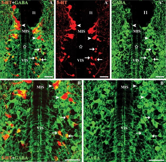
Fig. 3.
Transverse sections through the isthmic region of the rhombencephalon. (A-A″) Photomicrographs of a transverse section of the rostral rhombencephalon showing 5-HT-ir (red) and GABA-ir (green) cells. Note the presence of double-labelled cells in the ventral isthmus (white arrows) and the absence of double-labelled cells in the medial isthmic subgroup (arrowheads). (B-B′) Enlargements of Fig.2A-A″. Stars indicate the ventricle. Note that the anti-5-HT immunofluorescence labelling fills the somas, whereas the GABA labelling is more concentrated at the border of the neuronal somas. I1, Müller isthmic giant cell. All photomicrographs are Z-stack projections from confocal images. Scale bars: 50 µm.

