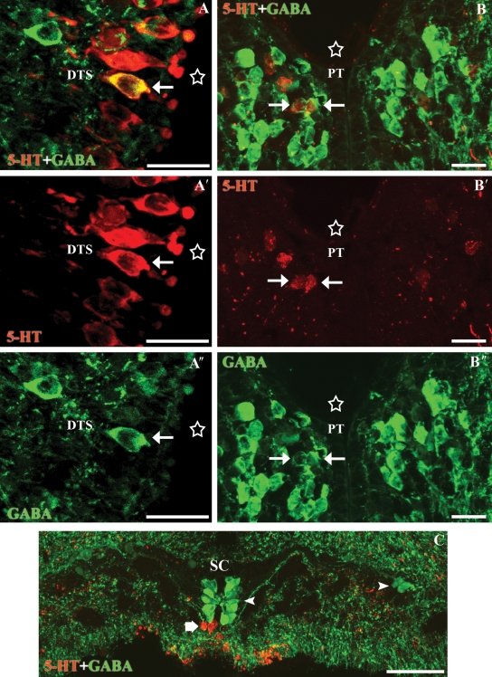
Fig. 4.
Transverse sections through the dorsal tuberal subgroup and the pretectal nucleus of the diencephalon and the spinal cord. (A-A″) Photomicrographs of a transverse section of the hypothalamus showing a 5-HT-ir (red)/GABA-ir (green) cerebrospinal fluid-contacting cell of the dorsal tuberal subgroup (white arrow). (B-B″) Photomicrographs of a transverse section of the caudal diencephalon showing a couple of double-labelled cells in the pretectal nucleus (white arrows). Note that the cells of the pretectum were faintly immunoreactive for the anti-5-HT antibody (red). (C) Photomicrograph of a transverse section of the spinal cord illustrating the absence of double-labelled cells (the thick arrow indicates 5-HT-ir cells and the arrowheads indicate GABA-ir cells). Stars indicate the ventricle. All photomicrographs are Z-stack projections from confocal images. Scale bars: (A) 20 µm, (B) 25 µm, (C) 40 µm.

