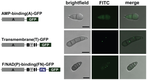
Figure 3. The transmembrane domain of the TmpL protein carries an organelle targeting signal.
Organelle targeting of partial or complete TmpL-GFP fusion proteins. At left schematic representations: gray boxes represent the AMP-binding (A) domain of TmpL protein; six tandem black boxes represent the transmembrane (T) domain; and a blue box represents the FAD/NAD(P)-binding (FN) domain. Right micrographs show GFP signal localization patterns of each fusion protein in A. brassicicola conidia. Bars = 10 µm.

