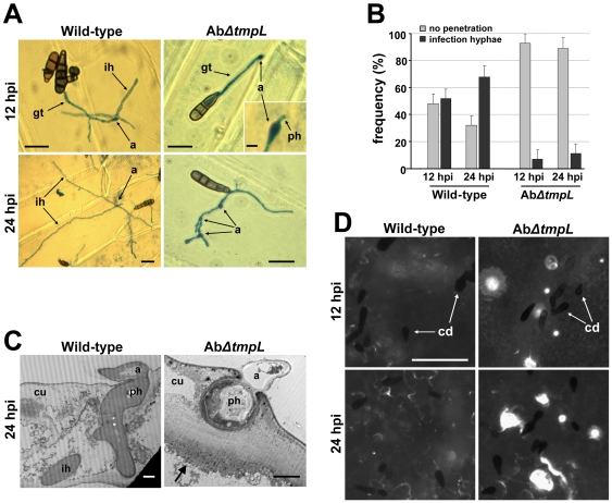
Figure 10. Appressoria and infection hyphae formation, ultrastructure, and callose detection assays of A. brassicicola ΔtmpL mutant infection.
(A) Light micrographs of onion epidermis inoculated with A. brassiciola wild-type and ΔtmpL mutants at 12 and 24 hpi. Note that penetration hyphae (inset) were frequently visible under the mutant appressoria, but no further development was observed in the AbΔtmpL mutant infection. Bars = 20 µm, except for the inset where it denotes 5 µm. Abbreviations: a, appressorium; gt, germ tube; ih, infection hypha; ph, penetration hypha. (B) Frequency of infection hyphae formation beneath appressoria of the A. brassicicola wild-type and ΔtmpL mutants on onion epidermis. Columns and error bars represent average and SD, respectively, of four independent experiments. (C) Transmission electron micrographs showing transverse sections of green cabbage leaves inoculated with A. brassicicola wild-type and ΔtmpL mutants. Leaves were collected at 24 hpi and prepared for transmission electron microscopy. Arrow indicates papilla formation (callose deposition) around a fungal penetration hypha. Bars = 2 µm. Abbreviations: a, appressorium; cu, cuticle layer; ih, secondary infection hypha; ph, penetration hypha. (D) Callose deposition on green cabbage cotyledons inoculated with A. brassicicola wild-type and ΔtmpL mutants. White spots indicate callose deposition of the inoculation sites stained by aniline blue and viewed by epifluorescence microscopy. The tiny black spots dispersed on plant surface are fungal conidia (cd). Bars = 50 µm.

