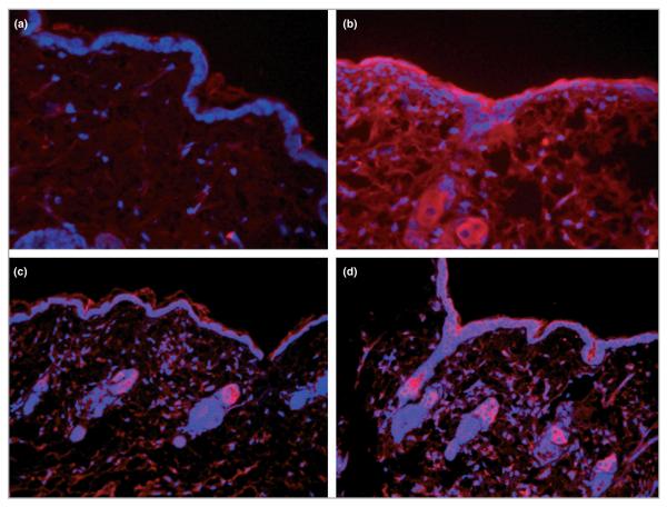
Fig 4.
Immunohistology of transforming growth factor (TGF)-β1 and TGF-β type 2 receptor (TGF-βR2) in intradermal injection skin samples. Interleukin (IL)-6 knockout (KO) mice were injected with 10 μL of 0 ng mL−1 (a, c) or 100 ng mL−1 (b, d) recombinant mouse (rm) IL-6, and mRNA was extracted from the tissue as described. Formalin-fixed, paraffin-embedded tissue sections of treated skin samples were incubated with anti-TGF-β1 (a and b), anti-TGF-βR2 (c and d) antibodies, and detected using Alexflor 546 conjugated secondary antibodies. Stained slides were visualized under fluorescence using a Leica DM400B microscope and 20 × objective.

