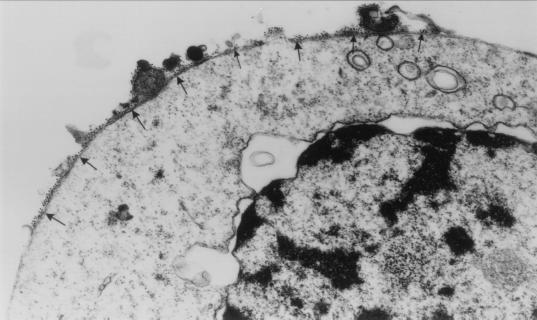
Figure 5.
Localization of the Porimin antigen by immunoelectron microscopy. Jurkat cells were labeled with anti-Porimin followed by goat anti-mouse Ig conjugated to 10-nm gold particles and examined by transmission electron microscopy (×35,000 original magnification). The gold particles are localized onto the surface of plasma membrane (arrows) of the cell.

