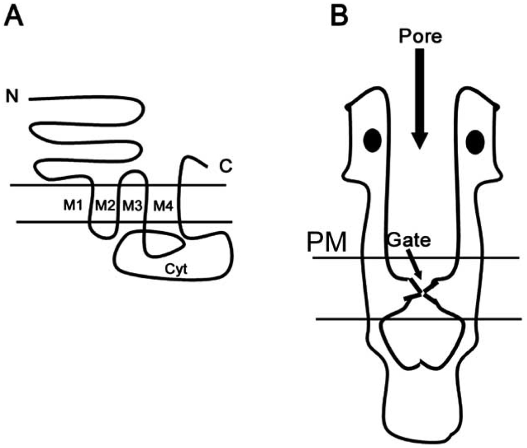
Fig. (3). Basic Structure of nAChRs.
A. The figure shows a typical 4 transmembrane topology of nAChRs from the N- to C- termini (N & C). Transmembrane domains are denoted (M1–M4). There is a large cytoplasmic loop separating M4 from M3 (cyt).
B. A low resolution model of nAChRs based on cryo-EM and X-ray crystallographic studies. There is a large N-terminal portion of the protein that forms the extracellular portion of the receptor. The distance between residues lining the ion channel pore opening narrows as the protein spans the plasma membrane. The narrowest portion of the pore lining region, lined by a ring of leucine residues, forms the channel gate. This is followed by the large cytoplasmic region. The two ligand-binding domains are depicted as filled ovals in the figure.

