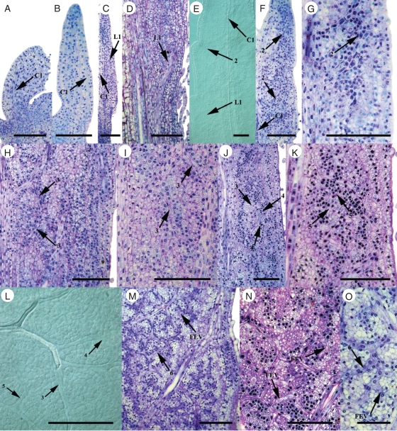
Fig. 4.
Onset and early development of vein orders in paradermal sections and clearings of leaves of C4 Flaveria bidentis. (A) Basal portion of young leaf showing central 1° vein procambium (0·5 mm). (B) Young leaf showing central 1° vein procambium (1 mm). (C) Mid-portion of leaf from central 1° vein to margin with lateral 1° vein procambium (1 mm). (D) Basal portion of leaf with lateral 1° vein procambium (3 mm). (E) Mid-portion of leaf with lateral 1° and 2° vein procambium (2 mm). (F) Leaf showing onset of 2° vein procambium (1 mm). (G) Connection of 2° vein loops (1 mm). (H) Appearance of 3° vein mesh (2 mm). (I) Developing 3° vein procambium within a 2° vein loop (2 mm). (J) 4° minor veins branching from 3° veins in basal portion of leaf (4·5 mm). (K) Serial section of leaf in J showing 5° minor veins. (L) Mid-portion of leaf with minor vein procambium (4·5 mm). (M) Mid-portion of the leaf with developing minor vein reticulum (7 mm). (N) Appearance of freely ending veinlets connected to minor veins in mid-portion of leaf (5 mm). (O) Mid-portion of leaf with freely ending veinlets compared with connected minor veins (7 mm). Abbreviations: C1, central 1° vein; L1, lateral 1° vein. Scale bars: all images = 100 µm, except (O) = 50 µm.

