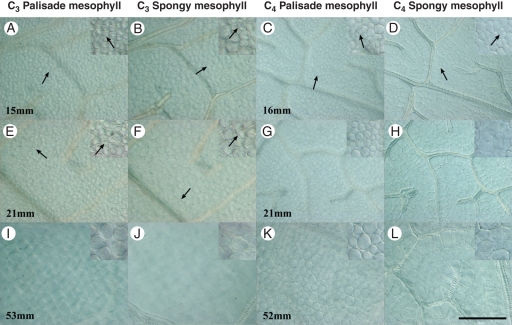
Fig. 8.
Palisade and spongy mesophyll development from clearings of C3 and C4 Flaveria leaves. Insets show enlargement and recent divisions of palisade and spongy mesophyll. (A, B, E, F, I, J) Mesophyll cells enlarge showing morphological and size differences between palisade and spongy mesophyll in C3 F. robusta. Arrows indicate recently divided cells. (C, D, G, H, K, L) Mesophyll cells enlarge showing little morphological and size differences between palisade and spongy mesophyll in C4 F. bidentis. Arrows indicate recently divided cells. Scale bar: all images = 100 µm; insets are 2×.

