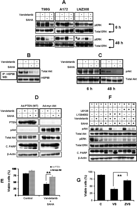
Fig. 8.
Combination of vandetanib and SAHA modulates survival and other regulatory molecules. A, logarithmically growing T98G, A172, and LNZ308 cells were incubated in the presence of 2 μM SAHA with or without vandetanib (2 μM) for different durations. Cells were lysed and 30 μg of total protein/lane was separated by SDS-PAGE and subjected to immunoblot analysis with phospho ERK 1/2 antibodies. Western blot analysis was performed as described under Materials and Methods. The blots were subsequently stripped and reprobed against total ERK. B, T98G cells were incubated in the presence of 2 μM SAHA with or without vandetanib (2 μM) for 48 h, after which HSP90 was immunoprecipitated (IP) from the cell lysates and immunoblotted (WB) with either anti-HSP90 or total Akt. Immunoprecipitation and Western blot analysis was performed as described under Materials and Methods. C, logarithmically growing T98G cells were incubated in the presence of 2 μM SAHA with or without vandetanib (2 μM) for the indicated durations. Cells were lysed and 30 μg of total protein/lane was separated by SDS-PAGE and subjected to immunoblot analysis with the phospho Akt (Ser 473) antibody. Western blot analysis was performed as described in Materials and Methods. The blots were subsequently stripped and reprobed against total Akt. D, logarithmically growing A172 cells were infected with wild-type PTEN or constitutively active form of Akt (Ad-myr-Akt) at 100 MOI per cell. Thirty-six hours after infection, cells were incubated in the presence of SAHA (2 μM) with or without vandetanib (2 μM) for 48 h. Control cells received DMSO. Cells were lysed and 30 μg of total protein/lane was separated by SDS-PAGE and subjected to immunoblot analysis with the indicated antibodies. E, logarithmically growing A172 cells were infected with wild-type PTEN (Ad-PTEN) or constitutively active form of Akt (Ad-myr-Akt) at 100 MOI per cell. Thirty-six hours after infection, cells were incubated in the presence of SAHA (2 μM) and vandetanib (2 μM) for 48 h. Control cells received DMSO. At the end of the incubation period, the viable cell numbers were determined by trypan-blue exclusion assay. For each analysis, at least 400 cells were evaluated in duplicate. The values represent the mean ± S.D. for two separate experiments performed in triplicate (∗∗, P < 0.005). F, logarithmically growing T98G cells were exposed to vandetanib (2 μM) or SAHA (2 μM) or the combination of both with or without U0126 or LY294002. Cells were pretreated with U0126 (5 μM) or LY294002 (5 μM) in complete medium 60 min before treatment with vandetanib and/or SAHA for 48 h. Western immunoblot analysis was performed with the indicated antibodies. G, T98G cells were pretreated with or without 50 μM Z-VAD-FMK (ZVS) in complete medium 60 min before treatment with vandetanib (2 μM) and SAHA (2 μM) (VS) for 48 h. Control cells received DMSO (C). At the end of the incubation period, the viable cell numbers were determined by trypan-blue exclusion assay. For each analysis, at least 400 cells were evaluated in duplicate. The values represent the mean ± S.D. for two separate experiments performed in triplicate (∗∗, P < 0.001).

