
Figure 1.
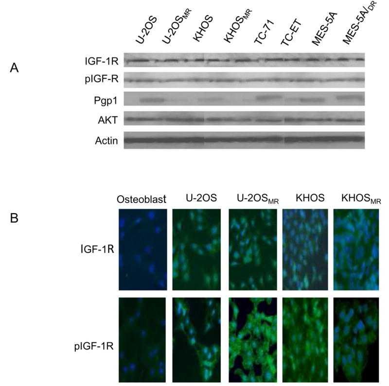
Expression of IGF-1R, phospho-IGF-1R (pIGF-1R) in the sarcoma cell lines as detected by Western blotting (A), immunofluorescence (B). A: Expression of IGF-1R, pIGF-1R in sarcoma drug sensitive and resistant cell line pairs determined by Western blot. Total cellular protein isolated from the indicated cell lines and immunoblotted with specific antibodies as described in Materials and Methods. The blots were also probed with an anti-actin monoclonal antibody to assess relative protein levels in the sample lanes. B: Confirmation of expression of IGF-1R, pIGF-R in osteosarcoma drug sensitive and resistant cell lines by immunofluroescence. Stained cells were then visualized on a Nikon Eclipse Ti-U fluorescence microscope.