Figure 7.
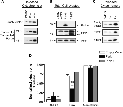
PINK1 does not prevent BH3 peptide-induced cytochrome c release (A) A representative western blot analysis of released cytochrome c release in a BH3 peptide assay following transient transfection to express parkin at 24 and 48 h. (B) Western blot analysis of whole cell lysates from MES cells lines transiently transfected with empty vector, wild-type parkin or PINK1. (C) Mitochondria were isolated 48 h after transient transfection with an empty vector control, parkin, or PINK1 and subject to a BH3 peptide assay. Western blot analysis of the Bim (10 µm) and alamethicin (Alm, 40 µg/ml) induced cytochrome c release demonstrates the effects of transiently expressed parkin and PINK1 on cytochrome c release. (D) Data from three independent experiments were analyzed by densitometry and normalized to the maximal cytochrome c release from each mitochondrial preparation (as determined by alamethicin treatment). *Denotes statistically significant from control (P < 0.05, n = 3) and † is significant from PINK1 (P < 0.05, n = 3).

