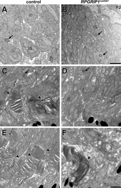
Figure 5.
Ultrastructural transmission electron microscopy analysis suggests normal CC formation and aberrant OS development. The CC (arrows) in (A) wild-type controls and (B) Rpgrip1nmf247 mutants did not differ (P2). At P8 OS discs are observed (arrow heads) in (C) wild-type controls but not in (D) Rpgrip1nmf247 mutants. At P9, OS are lengthened in (E) wild-type controls but (F) in the rare instance in which OS are observed in Rpgrip1nmf247 mutants, OS discs are enlarged and have a vertical orientation. IS, inner segment; RPE, retinal pigment epithelium. Bars = 500 nm.

