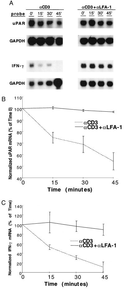
Figure 2.
Effect of LFA-1 engagement on T cell activation mRNA degradation. (A) Jurkat cells (for uPAR) or peripheral T cells (for IFN-γ) were treated with anti-CD3 or anti-CD3 plus anti-LFA-1 antibodies, panned, and incubated × 3 h at 37°C, after which transcription was arrested by the addition of DRB (0.2 mM). Antibody concentrations were as per Materials and Methods section, except that anti-CD3 0.5 μg/107 cells (five times standard protocol) were used to generate higher uPAR mRNA levels with anti-CD3 alone, allowing more easily interpretable decay curves. Total RNA was harvested at the indicated time points and used for uPAR, IFN-γ, and GAPDH Northern blots. Data displayed are representative of results from five separate experiments. Northern signals were densitometrically analyzed and displayed as % of maximal (time 0) GAPDH-normalized, densitometric units with mean values ± SE, for uPAR (B) and IFN-γ (C).

