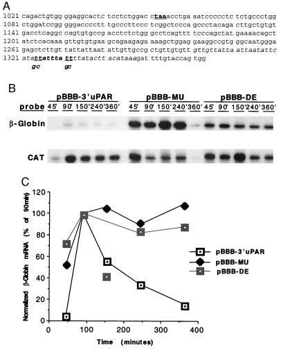
Figure 4.
Degradation of β-globin mRNA containing wild-type or mutant uPAR 3′ UTR. (A) The 3′ uPAR cDNA, with stop codon (taa) bold and underlined. Sequence 1084–1347 was subcloned into pBBB to generate pBBB-3′ uPAR. AU-rich region is italicized and deleted in pBBB-DE. The nonameric degradation motif is italicized and bolded. The underlined bases (t) in this sequence were mutated to g and c, as indicated, to generate pBBB-MU. (B) Jurkat cells were cotransfected with pEF-BOS-CAT and either pBBB 3′ uPAR, pBBB-MU, or pBBB-DE. Cells were fetal bovine serum-stimulated after a 24-h serum starvation, after which total RNA was harvested at the indicated time points for ribonuclease protection assay. Findings were similar in three separate experiments. (C) β-globin RPA signals from B were densitometrically analyzed and normalized to CAT signals. Isolated data point in the pBBB-DE curve (150 min) represents an aberrant experimental sample, not reproduced in other similar experiments.

