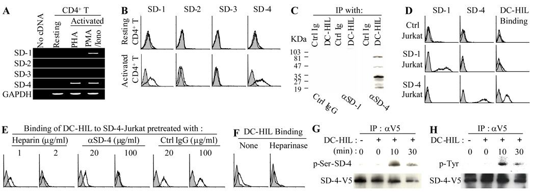
Figure 2. SD-4 is the T cell ligand of DC-HIL.
(A) mRNA expression of SD-1, -2, -3, -4, or GAPDH was examined by RT-PCR in total RNA isolated from resting CD4+ T cells or those activated with PHA or PMA/ionomycin. (B) Resting or PMA/ionomycin-activated CD4+ T cells were stained with Ab against SD-1, -2, -3, -4 (unshaded histograms) or control IgG (shaded), and surface expression examined by flow cytometry. (C) Whole cell extracts prepared from activated CD4+ T cells were immunoprecipitated with DC-HIL-Fc or control Ig and then immunoblotted with anti-SD-1, anti-SD-4, or control IgG. (D) Jurkat cells transfected with vector alone (Ctrl), SD-1 or SD-4 gene were examined by flow cytometry for surface expression of SD-1 or SD-4. These Jurkat transfectants were also treated with Con A and then examined by flow cytometry for binding of DC-HIL-Fc (unshaded hisotograms) or control Ig (shaded). (E and F) Binding of DC-HIL-Fc to Con A-activated SD-4+ Jurkat cells was performed in the presence of heparin at indicated concentrations. SD-4+ Jurkat cells were also pretreated with anti-SD-4 Ab or control IgG (E) or without (None) or with heparinase prior to binding to DC-HIL (F). (G and H) Phosphorylation of SD-4. Jurkat cells were transfected with SD-4-V5 gene and stimulated with Con A prior to incubation with immobilized DC-HIL-Fc. At varying time points after incubation, SD-4-V5 protein was immunoprecipitated with anti-V5 Ab, and serine (G) and tyrosine (H) phosphorylation assayed by immunoblotting with Ab to serine-phosphorylated SD-4 (p-Ser-SD-4) and to phosphorylated tyrosine (p-Tyr), respectively. In each precipitant, the amount of SD-4-V5 protein precipitated was examined by immunoblotting. All data shown are representative of at least 2 separate experiments.

