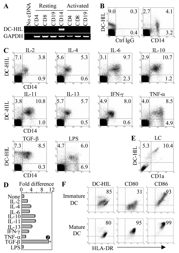
Figure 4. Expression of DC-HIL by human leukocytes.
(A) mRNA expression of DC-HIL or GAPDH was examined by RT-PCR of total RNA prepared from freshly isolated (resting) or activated CD4+, CD8+ T cells, CD19+ B cells, and CD14+ monocytes. (B) PBMC were immunostained with 3D5 anti-DC-HIL and anti-CD 14 mAb or isotype control IgG. Fluorescent labeling was examined by flow cytometry; quadro-analysis is shown. (C and D) Following stimulation with relevant cytokines or LPS for 2 d, PBMC were examined for surface expression of DC-HIL and CD14 by flow cytometry. Effects on surface expression of DC-HIL on CD14+ cells were assessed by fold differences calculated by mean fluorescence intensity on treated/untreated cells (None) (D) (mean ± SD, n = 3). * p<0.05 vs. None using Student’s t test. (E) Expression on epidermal LC. Epidermal cell suspensions were fluorescently stained with 3D5 mAb (PE-labeling) and anti-CD1a Ab (FITC-labeling), and surface expression was analyzed by flow cytometry. Dot-blot is shown, with frequency (%) of stained cells. (F) Immature and mature DC were doubly-stained with anti-HLA-DR and 3D5 mAb or Ab to either one of two activation markers (CD80 and CD86). All data shown are representative of 3 independent experiments.

