Fig. 4.
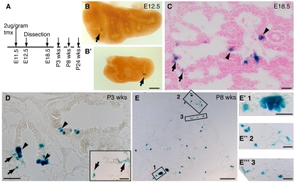
Single Id2+ tip epithelial cells can give rise to both bronchiolar and alveolar lineages. (A) Id2-CreERT2; Rosa26R-lacZ mouse embryos were exposed to a low dose of tmx at E11.5 and sacrificed at intervals. (B,B′) E12.5 wholemount lungs showing individual clones (arrows). Pairs of cells are frequently seen, suggesting that lineage-labeled cells divided prior to dissection. (C) E18.5 section. Note lineage-labeled bronchiolar (arrowheads) and alveolar (arrows) cells. (D) Lung section from a 3-week-old mouse. Note the larger groups of lineage-labeled cells. Inset shows labeled alveolar cells in the adjacent section. (E-E‴) Lung section from an 8-week-old mouse. Low-magnification image showing clone size. Box 1 shows a bronchiolar clone at higher magnification. Boxes 2 and 3 are high magnifications of alveolar regions showing labeled type 1 and 2 cells. Scale bars: 1 mm in B,E; 20 μm in C and insets in D,E′-E‴; 40 μm D.

