Figure 1.

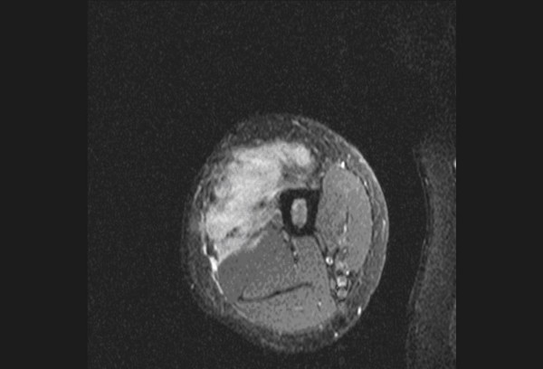
Post-contrast T1 fat saturated (FS) axial image of upper thigh shows irregular infiltrative margins of the malignant fibrous histiocytoma.

Post-contrast T1 fat saturated (FS) axial image of upper thigh shows irregular infiltrative margins of the malignant fibrous histiocytoma.