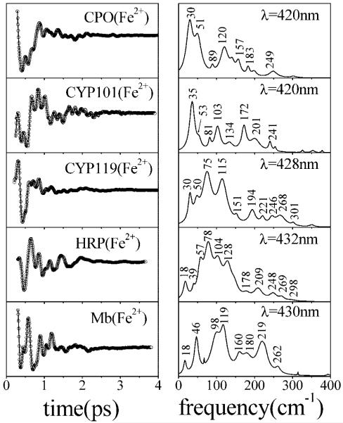
Figure 10.
Comparison of the low-frequency coherence spectra of three different cysteine-ligated ferrous heme proteins [CPO, P450CAM (CYP101) with camphor bound, and thermophyllic P450 (CYP119)] and two histidine-ligated ferrous heme proteins (reduced HRP and deoxymyoglobin). The left panels present the oscillatory signal and the LPSVD fit. The right panels show the corresponding coherence spectra. Experiments were carried out at wavelengths specified in the right panels using ~60 fs pulses.

