
Fig. 6.
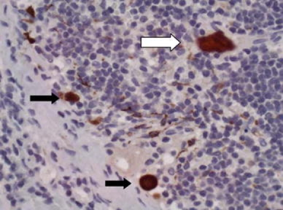
Cytokeratin-positive cells in a sentinel node stained by CKC pan cytokeratin. The white arrow shows a contaminant squame (this can be ascertained by the geometric outline, lack of nucleus, and by focusing at high power). The black arrows show nonnucleated individual tumor cells, and dendritic cells can be seen in the background