
Figure 4.
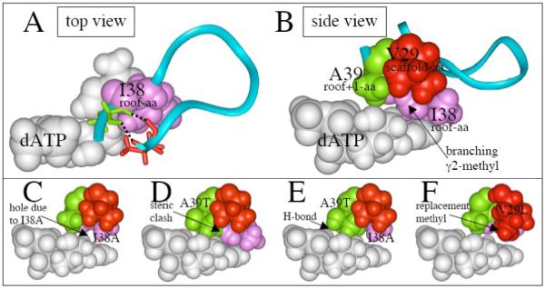
Structure of various regions of UmuC(V). (A) Top view showing how isoleucine-38 (purple) sits above dATP (white) in our UmuC(V) model, and is located in a loop (aa29-39) that includes V29 (scaffolding-aa, red), and A39 (roof+1-aa, green). The upper lip of this loop opens into the major groove, while the lower lip opens into a hole/cleft on the minor groove side, which can be analyzed based on analogy to a “chimney,”48 and which others call the “gap.”17 (B) Side view of the same structure as in panel A. (C) Model of UmuC(V) with isoleucine-38 converted to alanine-38 (I38A). (D) Model of UmuC(V) with alanine-39 converted to threonine-39 (A39T). (E) Model of UmuC(V) with both I38A and A39T. (F) Model of UmuC(V) with I38A and with valine-29 converted to isoleucine-29 (I38A/V29I).