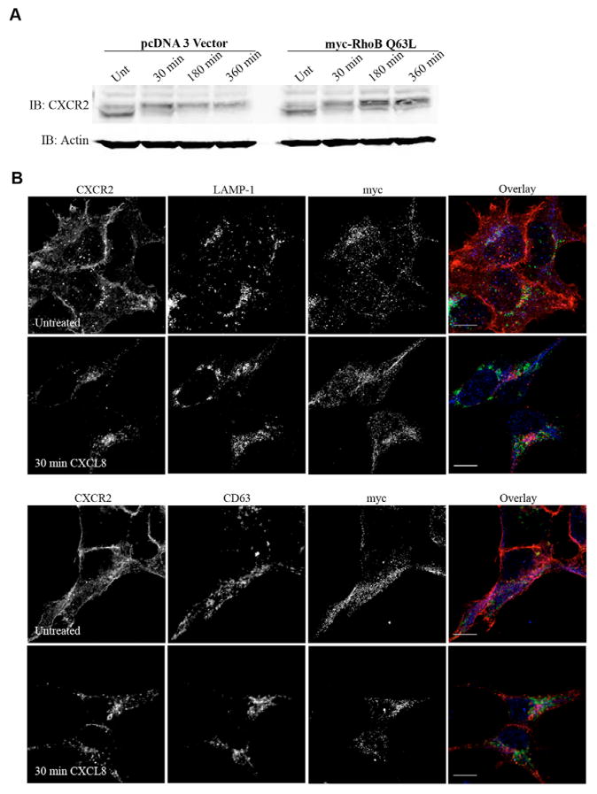
Fig. 8.
Expression of Myc-RhoB Q63L impairs CXCL8-induced CXCR2 degradation and does not result in colocalization with lysosomal markers. (A) Western blot analysis of CXCR2 expression in cells transfected with vector or Myc-RhoB Q63L in the presence of 20 μg/ml cycloheximide following stimulation with vehicle (Unt) or 100 ng/ml CXCL8 for 30 minutes, 180 minutes or 360 minutes. Actin is shown as a control to monitor for equal loading of protein. (B) Confocal images from three independent experiments of immunofluorescence-stained HEK293 cells stably expressing CXCR2 and transiently transfected with Myc-RhoB Q63L. Transfected cells were identified by staining with anti-Myc antibody. CXCR2 and LAMP-1 (top panels) or CD63 (bottom panels) staining in cells stimulated with vehicle (Untreated) or 100 ng/ml CXCL8 for 30 minutes. Overlay images are pseudocolored; red is CXCR2, green is LAMP-1 or CD63, blue is Myc-RhoB Q63L. Images were processed using Photoshop (Adobe Systems). Bars, 10 μm. Data shown are representative from three separate experiments.

