Abstract
RNA interference (RNAi) is the process of mRNA degradation induced by double-stranded RNA in a sequence-specific manner. Different types of promoters, such as U6, H1, tRNA, and CMV, have been used to control the inhibitory effect of RNAi expression vectors. In the present study, we constructed two shRNA expression vectors, respectively, controlled by tRNAlys and CMV enhancer-tRNAlys promoters. Compared to the vectors with tRNAlys or U6 promoter, the vector with a CMV enhancer-tRNAlys promoter silenced pokemon more efficiently on both the mRNA and the protein levels. Meanwhile, the silencing of pokemon inhibited the proliferation of MCF7 cells, but the induction of apoptosis of MCF7 cells was not observed. We conclude that the CMV enhancer-tRNAlys promoter may be a powerful tool in driving intracellular expression of shRNA which can efficiently silence targeted gene.
1. Introduction
RNAi was first discovered in the nematode Caenorhabditis elegant [1], after which the power and utility of RNAi for specifically gene silencing has driven its incredibly rapid adoption as a tool for reverse genetics at the organismal level. In mammalian cultured cells, RNAi is typically induced by siRNA introduced directly or expressed as a hairpin structure from a DNA construct, such as a plasmid, a viral vector, or a PCR product. It is known that the choice of promoters has been shown to be very important for efficient RNAi [2–4] and thus received more and more concern in vector-based expression systems after the introduction of vectors [4].The promoters U6 and H1 [5–15], which belong to Pol III, are widely used for inducible knock-down of gene expression in vitro or in vivo [16]. Type II Pol III promoters such as tRNA lead to longer primary dsRNAs than U6 or H1, which might induce interferon response that could be avoided by a careful design of vector system [6, 17, 18]. It has been reported that tRNA promoters are more effective in target gene knock down [6, 17]. Recently, a polymerase I promoter with high species specific has also been reported, which provides an interesting potential regarding the biosafety of experiments [19].
The CMV enhancer has been used in many vectors since it could increase the downstream DNA element's transcription level [20–22]. Recent studies have compared the inhibition of exogenous SOD1G93AGFP expression by CMV enhancer with U6 promoter and U6 promoter alone, and the results have shown a more efficient inhibition by the use of CMV enhancer [5]. However, silencing by enhancer-promoter constructs has not been compared with promoters alone for endogenous genes.
Pokemon is a POK protein family member, a specific ARF suppressor, whose expression level is a critical determinant of cellular response to oncogenic transformation, Pokemon acts as a genuine proto-oncogene when overexpressed [23]. It is thought that downregulated Pokemon could upregulate ARF, thus blocking MDM2, followed by the increase in p53 expression, and p53's activation may lead to apoptosis [24].
In this study, we constructed two kinds of shRNA expression vectors controlled, respectively, by tRNAlys and CMV enhancer-tRNAlys, in order to examine their efficiency. We then use the best shRNA to knock-down Pokemon expression and to observe the subsequent effect.
2. Materials and Methods
2.1. Plasmid Construction
The human tRNAlys promoter was PCR-amplified from genomic DNA isolated from HeLa cells. Primers used for the generation of tRNAlys were P(Lys1): 5′-GAT GAT ATC TGG CCA CTA GGG ACT GGG-3′, and P(Lys2): 5′-GTC GGA TCC AAG CTT GAA TTC GGG CCC AGT CTG ATG CTC TAC CGA CTG-3. The PCR product was digested with BamHI and EcoRV and then named tRNAlys promoter.
The plasmid backbone with CMV enhancer was PCR-amplified from plasmid pCMV5. Primers used were P(MRV1): 5′-GTG GAT ATC GGG GCG GGG TTA TTA CGA C-3′, and P(MRV2): 5′-CGA CTC TAG AGG ATC CCG GGT G-3′. The PCR product was digested with BamHI and EcoRV and then named pCBE. Through ligation of tRNAlys promoter and pCBE, a novel vector was created and thus designated as pExsiler, which derived from pCMV5 through replacing CMV minimal promoter with tRNAlys promoter.
In order to obtain the plasmid containing one tRNAlys promoter without CMV enhancer, the pCMV5 plasmid was first digested with BamHI, EcoRI, followed by HindII to completely remove the CMV enhancer sequence and CMV minimal promoter. The processed plasmid fragment was named pBE. Through ligation of tRNAlys promoter and pBE, a new vector was created and thus designated as pSiler, which derived from pCMV5 through replacing CMV promoter (CMV enhancer and CMV minimal promoter) with tRNAlys promoter.
The fragment of human Pokemon cDNA was PCR-amplified from cDNA isolated from MCF7 cells. Primers used in this PCR were P(POK1): 5′-CTTAAGCTTGCCACCATGGCCGGCGGCGTGG-3′, and P(POK2): 5′-CATGATATCGGCGAGTCCGGCTGTGAAGTTAC-3′. The PCR product was digested with HindIII and EcoRV and then cloned into the HindIII/SmaI sites of pEGFP(N2). The recombinant plasmid was named pEGFP(N2)-Pokemon. SiRNA target sequences of human Pokemon gene were designed using the Takara siRNA Design Support System (http://www.takara.com.cn/enter.htm). Among the 10 target sequences identified by this free online software,three target sequences were selected as follows: (1) CGTGTACGAGATCGACTTC; (2) CGGCTTAGACTTCTATGGG; (3) GGGCGTCATGGACTACTAC. According to these three target sequences, the sense strands and antisense strands of duplex shRNA oligonucleotides were synthesized as shown in Table 1.
Table 1.
Sequences of oilgonucleotides used in the research.
| shRNA | Sequences of oilgonucleotides | Target site on pokemon mRNA (GenBank accession number NM_01 5898) |
|---|---|---|
| 1 | F1: 5′-ccgtgtacgagatcgacttcttcaagagagaagtcgatctcgtacacgtttttt-3′ | 383–401 nt |
| R1: 3′-ccggggcacatgctctagctgaagaagttctctcttcagctagagcatgtgcaaaaaatcga-5′ | ||
| 2 | F2: 5′-ccggcttagacttctatgggttcaagagacccatagaagtctaagccgtttttt-3′ | 809–827 nt |
| R2: 3′-ccggggccgaatctgaagatacccaagttctctgggtatcttcagattcggcaaaaaatcga-5′ | ||
| 3 | F3: 5′-cgggcgtcatggactactacttcaagagagtagtagtccatgacgccctttttt-3′ | 1202–1200 nt |
| R3: 3′-ccgggcccgcagtacctgatgatgaagttctctcatcatcaggtactgcgggaaaaaatcga-5′ |
shRNA encoding cassettes 1–3 were formed by annealing two corresponding oligonucleotides, respectively. F stands for forward and R for reverse.
These single strand oligonucleotides were phosphorylated, and then F1 and R1, F2 and R2, and F3 and R3 oligonucleotides were annealed to generate the duplex oligonucleotides named as FR1, FR2, and FR3. FR1, FR2, and FR3 duplex oligonucleotides were cloned into the ApaI/HIndIII sites of pExsiler and pSiler, and the resulting plasmids were named EP1, EP2, EP3, SP1, SP2, and SP3, respectively. FR3 double-stranded oligonucleotides were cloned into the ApaI/HIndIII sites of pBS/U6(kindly provided by Yang Shi of Harvard University) to create the plasmid designated as UP3.
In order to obtain a control plasmid with only TTTTTT sequence at the shRNA site, a PCR was preformed, using P(WW1): 5′-GTC GAT ATC AGA TCT TGA CAT TGA TTA TTG ACT AGT TAT TAA TAG 3′, and P(WW2): 5′-GTC GGA TCC AAA AA AGG GCC CAG TCT GAT GCT CTA CCG ACT G-3′ as primers and pExsiler as the template. The PCR product was digested with BamHI and EcoRV and then cloned into pBE. The resulting plasmid was named ET as a control plasmid.
2.2. Cell Culture and Transfection of Cells
Human galactophore cancer cell line MCF-7 was cultured in Dulbecco's modified Eagle's medium (DMEM) (Invitrogen) supplemented with 10% fetal bovine serum (FBS) (Hyclone), 2 mM glutamine (Invitrogen), 100 U/mL penicillin, and 100 mg/mL streptomycin at 37°C in a humidified 5%CO2 incubator.
For western blot and the detection of apoptosis, twenty-four hours before transfection, 5 × 10⁵ MCF-7 cells were trypsinized and transferred to 6-well plates and cultured in DMEM supplemented with 10% FBS without antibiotics. They were grown to a confluency of 90%–95% and then transfected with a total of 4 μg of each of the hairpin vectors using lipofectamine 2000 (Invitrogen) according to the manufacturer's protocol, except that the transfection volume was reduced to 1.5 mL in order to obtain higher transfection efficiency. For the screening of the RNAi plasmids, separate pEGFP(N2)-Pokemon plasmid encoding Pokemon-GFP fusion protein and the RNAi plasmids were generally used at a ratio of 1 : 1 [16]. Exogeneous GFP-Pokemon protein expression levels of EGFP were assessed by green fluorescent signals expression under a microscope. For the detection of cell proliferation, 1 × 10⁴ MCF-7 cells were trypsinized and transferred to 96 well plates, and cultured in DMEM supplemented with 10% FBS without antibiotics. They were immediately transfected with 0.2 μg of each of the hairpin vectors using lipofectamine 2000 (Invitrogen) according to the manufacturer's protocol.
2.3. Real Time RT-PCR
36 hours after transfection, MCF-7 cells were collected and washed three times with cold phosphate buffered saline (PBS). Total cellular RNA was prepared using Trizol reagent (Invitrogen). 1 μg of total RNA was reverse transcribed (RT) using Superscript II (Invitrogen) and random hexamers according to the manufacturer's protocol. 1 μL cDNA was added to 19 μL RealMastermix (SYBR Green) (TIANGEN) containing 1x RealMasterMix, 1x SYBR Green (TIANGEN), 200 nM of sense primer FW (5′- GAA GCC CTA CGA GTG CAA CAT C-3′), and 200 nM of antisense primer ReV (5′-GTG GTT CTT CAG GTC GTA GTT GTG -3′) [23]. Real time PCR was carried out in an ABI Appied Biosystem 7500 (Applied Biosystems) using the following thermal cycling profile: 94°C 1 minute, followed by 40 cycles of amplification (94°C 20 seconds, 60°C 20 seconds, 68°C 40 seconds). For RNA normalization, a GAPDH PCR was performed with the same PCR reagents except that the primers were G1 (5′- GGT GGT CTC CTC TGA CTT CAA CA-3′) and G2( 5′-GTT GCT GTA GCC AAA TTC GTT GT-3′) [25].
2.4. Western Blot
MCF-7 cells were collected 48 hours after transfection. Cells were washed with cold PBS three times and collected in 500 μL PBS using a cell scraper and transferred into an Eppendorf tube. Following centrifugation, cells were resuspended in PBS. Cells were lysed in TNE lysis buffer (10 mM Tris, pH 7.8, 150 mM NaCl, 1 mM EDTA, 0.5% NP-40) containing protease inhibitor PMSF. After 30-minutes incubation on ice, the lysates were spun at 12 000 g for 15 minutes to remove insoluble material. Protein concentrations were determined through the Bradford procedure. 20 μg of total protein lysates were resolved on a 12% SDS-PAGE gel and transferred to a Nitrocellulose membrane (Life sciences). Membranes were blocked with 5% nonfat dried milk powder in TBS/T (0.05% Tween-20) for 2 hours at room temperature. Membranes were sequentially incubated with LRF (E-15) (Santa Cruz sc-34510) and donkey antigoat IgG-HRP (Santa Cruz sc-2020) according to the manufacture's protocol. The protein bands were visualized using SuperSignal West Pico Chemiluminescent Substrate (PIERCE) according to the manufacture's protocol.
For sequential probing, membranes were stripped in Erasure buffer (2% SDS, 62.5 mM Tris-HCl PH 6.8, 100 mM beta-mercaptoethanol) at 70°C for 40 minutes and then incubated in TS buffer (10 mM Tris-HCl PH 7.4, 150 mM NaCl). Actin bands were then detected using polyclone antibody rabbit anti β-actin PR-0255 (ZHONGSHAN GOLDBRIDGE, Beijing, China) and HRP labeled antibody goat antirabbit ZDR-5306 (ZHONGSHAN GOLDBRIDGE).
2.5. Detection of Cell Proliferation
The proliferation rate of cell line MCF-7 was measured with a Cell proliferation Kit II (Roche Ltd, Switzerland) according to the manufacturer's protocol.
2.6. Flow Cytometric Determination of Apoptosis
60 hours after transfection, MCF-7 cells were trypsinized and resuspended in PBS. Cells from 2 wells of a 6-well plate were collected and centrifuged, and the pellets were used for the flow cytometric determination of apoptosis using ANNEXIN V-FIFC APOPTOSIS DETECTION KIT I (BD biosciences) according to the manufacture's protocol. Cells treated with 10 μg/mL cisplatin served as a positive control.
3. Results
3.1. Construction of Plasmids
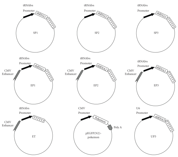
To compare the efficiencies of CMV enhancer-tRNAlys promoter and tRNAlys promoter, we constructed two kinds of expression vectors (pExsiler and pSiler )and attached dsRNA to the 3′ end of each promoter. All plasmids (pExsiler, pSiler, pEGFP(N2)-Pokemon, EP1, EP2, EP3, SP1, SP2, SP3, UP3, and ET) were sequenced (Invitrogen, China). Results of sequencing verified that the sequences of the all aforementioned plasmids were consistent with what we had designed (Figure 1).
Figure 1.
Map of construscts used in this work. SP1, SP2 and SP3 plasmids derived from the pSiler plasmid can express shRNA1, shRNA2, and shRNA3, respectively. EP1, EP2 and EP3 plasmids derived from the pExsiler plasmid can express shRNA1, shRNA2, and shRNA3, respectively. UP3 plasmid derived from pBS/U6 plasmid can express shRNA3. As control, ET plasmid derived from the pExsiler plasmid can express no shRNA. pEGFP(N2)-Pokemon derived from pEGFP-N2 vector can express Pokemon-GFP fusion protein.
3.2. Efficiency of RNAi between Different shRNA Expression Plasmids
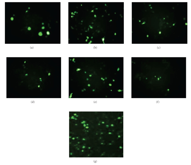
For selection of most efficient shRNA from the three shRNA sequences of human Pokemon gene, the EP1, EP2, EP3, SP1, SP2, SP3, and ET plasmids were respectively, cotransfected together with the pEGFP(N2)-Pokemon into MCF7 cells. Pokemon-GFP expression within the transfected cells was assayed by green fluorescent signals (Figure 2). Whereas EP2 and SP2 had less effect on Pokemon-GFP expression, EP3 and SP3 greatly diminished its expression. Therefore, the shRNA derived from target sequences (GGGCGTCATGGACTACTAC) has most efficiency in knockdown of Pokemon expression.
Figure 2.
Expression of Pokemon-GFP in MCF7 cells cotransfected with the pEGFP(N2)-Pokemon and different shRNA expression plasmids. (a) MCF7 cells cotransfected with the pEGFP(N2)-Pokemon and SP1, (b) with the pEGFP(N2)-Pokemon and SP2, (c) with the pEGFP(N2)-Pokemon and SP3, (d) with the pEGFP(N2)-Pokemon and EP1, (e) with the pEGFP(N2)-Pokemon and EP2, (f) with the pEGFP(N2)-Pokemon and EP3, and with the (g) pEGFP(N2)-Pokemon and ET.
3.3. Efficiency of RNAi between Different Expression Plasmids
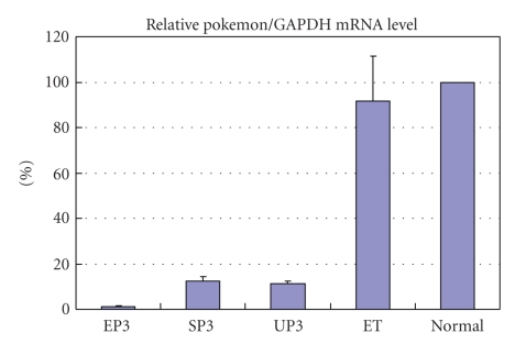
To examine the efficiency of RNAi in MCF-7 cells, we introduced expression plasmids EP3/SP3/UP3/ET into MCF-7 cells which express Pokemon. We examined Pokemon mRNA levels through real time RT-PCR. As shown in Figure 3, there is no obvious decrease of Pokemon/GAPDH mRNA level in cells transfected with ET. However, cells transfected with EP3, SP3, and UP3 show decreased relative Pokemon/GAPDH mRNA levels to 1.2% ± 0.2%, 12.3% ± 2.2%, and 11.1% ± 1.3%, respectively, compared to normal MCF-7 cells.
Figure 3.
Efficiency of different shRNA constructs in the inducing of silencing on Pokemon/GAPDH mRNA level as quantified by real-time RT-PCR. Total RNA was extracted from MCF-7 cells 36 hours after transfection with the indicated constructs. Normal MCF-7 cells were used as a negative control. An external standard curve was created to make sure that Pokemon and GAPDH were amplified at similar efficiency. Data shown represents, three replicated experiments with error bar indicated.
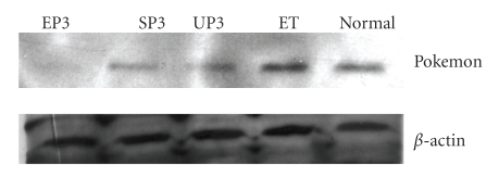
Western blot was used to examine the Pokemon expression level. Figure 4 shows that the Pokemon expression level in MCF7 cells transfected with EP3 was significantly lower than those with SP3, UP3 and ET, whereas the β-actin expression levels remained unchanged. These results suggest that the enhancer-promoter construct can specifically and efficiently silence Pokemon on both mRNA and protein levels in MCF-7 cells.
Figure 4.
Efficiency of different shRNA constructs in the inducing of silencing of endogenous gene expression. The western blot shows the Pokemon bands and the β-actin bands which serve as an endogenous control.
3.4. Effects of Pokemon Inhibition
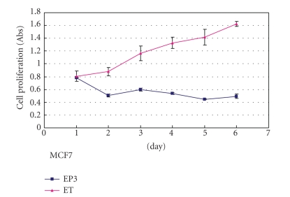
To examine the phenotype of cells that were transfected with different constructs, the proliferation rates of MCF-7 were analyzed. As shown in Figure 5, MCF-7 cells transfected with EP3, in which Pokemon levels were reduced significantly both on mRNA and protein scale, proliferated much more slowly than cells transfected with ET, which was served as a negative control. The reduction of proliferation rate is believed to be at least partially due to the decrease of Pokemon protein level in MCF-7 cells.
Figure 5.
Effect of proliferation of cells transfected with different shRNA constructs. Rates of proliferation were determined as described in the text. Data are mean values from three replicates with error bar indicated. MCF-7 cells transfected with EP3 proliferateare significantly more slowly than cells transfected with control plasmid ET. The wild type MCF-7 cells were not shown.
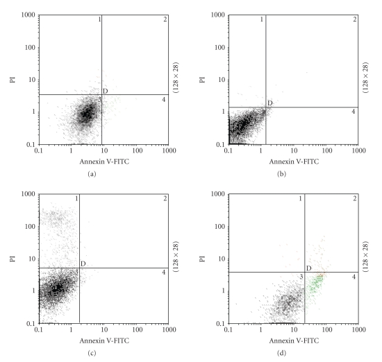
Recent research has suggested that the inhibition of Pokemon might result in the apoptosis or the cell cycle arrest [23]. However, in the present study, no typical apoptosis was observed by Flow Cytometry in MCF-7 cells transfected by EP3 and ET, while cells treated with cisplatin did show apoptosis (Figure 6).
Figure 6.
Flow cytometric determination of apoptosis. (a) Unstained normal MCF-7 cells (b) EP3-transfected cells stained with both PI and Annexin V-FITC (c) ET-transfected cells stained with both PI and Annexin V-FITC (d) Cisplatin-treated cells stained with both PI and Annexin V-FITC. Note that some cells transfected with PT were dead as a result of high cell density. Results are representative of two replicates.
4. Discussions
In the present study, we found that CMV enhancer, one of the widely used enhancers, could increase the efficiency of tRNAlys promoter. It has been reported that the communication between the enhancer and the promoter could induce gene activation [26], while the disruption of the communication leads to the inhibition of gene expression [27]. Therefore, enhancers may activate the transcription of downstream shRNA, resulting in the increase of RNAi efficiency.
Pokemon, also known as LRF, OCZF, and FBI-1, is a POK protein family member, a specific ARF suppressor, whose expression level is a critical determinant of cellular response to oncogenic transformation. So Pokemon acts as a genuine proto-oncogene when over-expressed [23]. It is thought that downregulated Pokemon results in an increase in p53 expression, and p53's activation may lead to apoptosis [24]. In our study, no apoptosis but the slower proliferation rate is observed in the inhibition of Pokemon in MCF-7 cells induced by shRNA controlled by CMV enhancer with tRNAlys promoter. However, as the ARF/p53 pathway is frequently abrogated in human cancers, if MCF-7 cells do lose this path, this apparent discrepancy could be solved. What is more, current evidences show that Pokemon null mouse resulted in embryonic lethal due to severe anemia and profoundly impaired differential cellular in multiple tissues [23]. But MCF-7 cells are already differentiated cells; thereby they may avoid apoptosis for that reason. However, more investigation should be made to confirm our findings.
In conclusion, the shRNA expression vector controled by CMV enhancer-tRNAlys promoter is powerful in silencing Pokemon and may be a useful tool for scientific research and therapy use with RNAi.
Acknowledgments
The authors gratefully acknowledge the National Natural Science Foundation of China (NSFC20132020, 20572060) and the Ministry of Science and Technology of China (2007AA02Z160) for financial support of this work. The pBS/U6 plasmid was kindly provided by Yang Shi of Harvard University. they also thank Dr. Tan Chuanyan and Mr. Yan Dongdong for the manuscript.
References
- 1.Guo S, Kemphues KJ. par-1, a gene required for establishing polarity in C. Elegans embryos, encodes a putative Ser/Thr kinase that is asymmetrically distributed. Cell. 1995;81(4):611–620. doi: 10.1016/0092-8674(95)90082-9. [DOI] [PubMed] [Google Scholar]
- 2.Sohail M, Doran G, Riedemann J, Macaulay V, Southern EM. A simple and cost-effective method for producing small interfering RNAs with high efficacy. Nucleic Acids Research. 2003;31(7):p. e38. doi: 10.1093/nar/gng038. [DOI] [PMC free article] [PubMed] [Google Scholar]
- 3.Donzé O, Picard D. RNA interference in mammalian cells using siRNAs synthesized with T7 RNA polymerase. Nucleic Acids Research. 2002;30(10):p. e46. doi: 10.1093/nar/30.10.e46. [DOI] [PMC free article] [PubMed] [Google Scholar]
- 4.Bertrand E, Castanotto D, Zhou C, et al. The expression cassette determines the functional activity of ribozymes in mammalian cells by controlling their intracellular localization. RNA. 1997;3(1):75–88. [PMC free article] [PubMed] [Google Scholar]
- 5.Xia XG, Zhou H, Ding H, el Affar B, Shi Y, Xu Z. An enhanced U6 promoter for synthesis of short hairpin RNA. Nucleic Acids Research. 2003;31(17):p. e100. doi: 10.1093/nar/gng098. [DOI] [PMC free article] [PubMed] [Google Scholar]
- 6.Boden D, Pusch O, Lee F, Tucker L, Shank PR, Ramratnam B. Promoter choice affects the potency of HIV-1 specific RNA interference. Nucleic Acids Research. 2003;31(17):5033–5038. doi: 10.1093/nar/gkg704. [DOI] [PMC free article] [PubMed] [Google Scholar]
- 7.Yu J-Y, DeRuiter SL, Turner DL. RNA interference by expression of short-interfering RNAs and hairpin RNAs in mammalian cells. Proceedings of the National Academy of Sciences of the United States of America. 2002;99(9):6047–6052. doi: 10.1073/pnas.092143499. [DOI] [PMC free article] [PubMed] [Google Scholar]
- 8.Paddison PJ, Caudy AA, Bernstein E, Hannon GJ, Conklin DS. Short hairpin RNAs (shRNAs) induce sequence-specific silencing in mammalian cells. Genes and Development. 2002;16(8):948–958. doi: 10.1101/gad.981002. [DOI] [PMC free article] [PubMed] [Google Scholar]
- 9.Czauderna F, Santel A, Hinz M, et al. Inducible shRNA expression for application in a prostate cancer mouse model. Nucleic Acids Research. 2003;31(21):p. e127. doi: 10.1093/nar/gng127. [DOI] [PMC free article] [PubMed] [Google Scholar]
- 10.Kwak Y-D, Koike H, Sugaya K. RNA interference with small hairpin RNAs transcribed from a human U6 promoter-driven DNA vector. Journal of Pharmacological Sciences. 2003;93(2):214–217. doi: 10.1254/jphs.93.214. [DOI] [PubMed] [Google Scholar]
- 11.Tiscornia G, Tergaonkar V, Galimi F, Verma IM. CRE recombinase-inducible RNA interference mediated by lentiviral vectors. Proceedings of the National Academy of Sciences of the United States of America. 2004;101(19):7347–7351. doi: 10.1073/pnas.0402107101. [DOI] [PMC free article] [PubMed] [Google Scholar]
- 12.Coumoul X, Shukla V, Li C, Wang RH, Deng CX. Conditional knockdown of Fgfr2 in mice using Cre-LoxP induced RNA interference. Nucleic Acids Research. 2005;33(11):p. e102. doi: 10.1093/nar/gni100. [DOI] [PMC free article] [PubMed] [Google Scholar]
- 13.Li M, Rohrer B. Gene silencing in Xenopus laevis by DNA vector-based RNA interference and transgenesis. Cell Research. 2006;16(1):99–105. doi: 10.1038/sj.cr.7310013. [DOI] [PubMed] [Google Scholar]
- 14.Brummelkamp TR, Bernards R, Agami R. A system for stable expression of short interfering RNAs in mammalian cells. Science. 2002;296(5567):550–553. doi: 10.1126/science.1068999. [DOI] [PubMed] [Google Scholar]
- 15.Seibler J, Küter-Luks B, Kern H, et al. Single copy shRNA configuration for ubiquitous gene knockdown in mice. Nucleic Acids Research. 2005;33(7):p. e67. doi: 10.1093/nar/gni065. [DOI] [PMC free article] [PubMed] [Google Scholar]
- 16.Pebernard S, Iggo RD. Determinants of interferon-stimulated gene induction by RNAi vectors. Differentiation. 2004;72(2-3):103–111. doi: 10.1111/j.1432-0436.2004.07202001.x. [DOI] [PubMed] [Google Scholar]
- 17.Kawasaki H, Taira K. Short hairpin type of dsRNAs that are controlled by tRNA(Val) promoter significantly induce RNAi-mediated gene silencing in the cytoplasm of human cells. Nucleic Acids Research. 2003;31(2):700–707. doi: 10.1093/nar/gkg158. [DOI] [PMC free article] [PubMed] [Google Scholar]
- 18.Oshima K, Kawasaki H, Soda Y, Tani K, Asano S, Taira K. Maxizymes and small hairpin-type RNAs that are driven by a tRNA promoter specifically cleave a chimeric gene associated with leukemia in vitro and in vivo. Cancer Research. 2003;63(20):6809–6814. [PubMed] [Google Scholar]
- 19.Brenz Verca MS, Weber P, Mayer C, et al. Development of a species-specific RNA polymerase I-based shRNA expression vector. Nucleic Acids Research. 2007;35(2):p. e10. doi: 10.1093/nar/gkl1045. [DOI] [PMC free article] [PubMed] [Google Scholar]
- 20.Su J, Zhu Z, Xiong F, Wang Y. Hybrid cytomegalovirus-U6 promoter-based plasmid vectors improve efficiency of RNA interference in zebrafish. Marine Biotechnology. 2008;10(5):511–517. doi: 10.1007/s10126-008-9087-8. [DOI] [PubMed] [Google Scholar]
- 21.Sawicki JA, Morris RJ, Monks B, Sakai K, Miyazaki J-I. A composite CMV-IE enhancer/beta-actin promoter is ubiquitously expressed in mouse cutaneous epithelium. Experimental Cell Research. 1998;244(1):367–369. doi: 10.1006/excr.1998.4175. [DOI] [PubMed] [Google Scholar]
- 22.Yew NS, Wysokenski DM, Wang KX, et al. Optimization of plasmid vectors for high-level expression in lung epithelial cells. Human Gene Therapy. 1997;8(5):575–584. doi: 10.1089/hum.1997.8.5-575. [DOI] [PubMed] [Google Scholar]
- 23.Maeda T, Hobbs RH, Merghoub T, et al. Role of the proto-oncogene Pokemon in cellular transformation and ARF repression. Nature. 2005;433(7023):278–285. doi: 10.1038/nature03203. [DOI] [PubMed] [Google Scholar]
- 24.Maeda T, Hobbs RM, Pandolfi PP. The transcription factor Pokemon: a new key player in cancer pathogenesis. Cancer Research. 2005;65(19):8575–8578. doi: 10.1158/0008-5472.CAN-05-1055. [DOI] [PubMed] [Google Scholar]
- 25.Lai J-P, Ho WZ, Kilpatrick LE, et al. Full-length and truncated neurokinin-1 receptor expression and function during monocyte/macrophage differentiation. Proceedings of the National Academy of Sciences of the United States of America. 2006;103(20):7771–7776. doi: 10.1073/pnas.0602563103. [DOI] [PMC free article] [PubMed] [Google Scholar]
- 26.Hatzis P, Talianidis I. Dynamics of enhancer-promoter communication during differentiation-induced gene activation. Molecular Cell. 2002;10(6):1467–1477. doi: 10.1016/s1097-2765(02)00786-4. [DOI] [PubMed] [Google Scholar]
- 27.Hatzis P, Kyrmizi I, Talianidis I. Mitogen-activated protein kinase-mediated disruption of enhancer-promoter communication inhibits hepatocyte nuclear factor 4alpha expression. Molecular and Cellular Biology. 2006;26(19):7017–7029. doi: 10.1128/MCB.00297-06. [DOI] [PMC free article] [PubMed] [Google Scholar]


