
Figure 1.
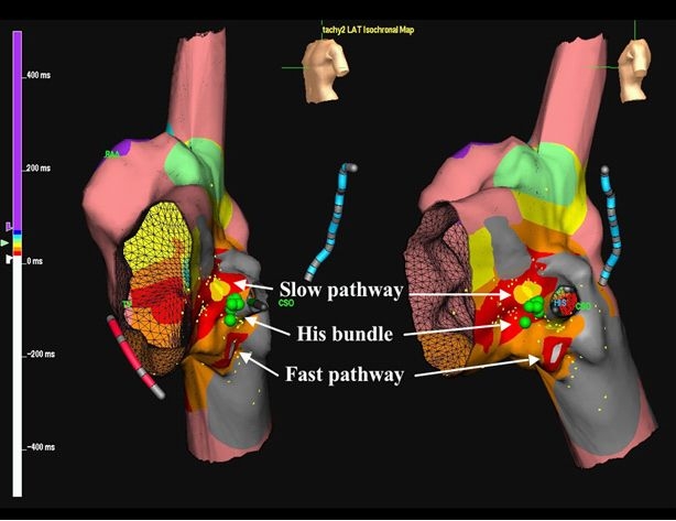
Cryomapping and cryoablation combined with 3D electroanatomic mapping in a patient with a partial AVCD. Three-dimensional electroanatomic maps of retrograde atrial activation during AV nodal reentrant tachycardia in left anterior oblique (Panel A) and left lateral (Panel B) views. The blue decapolar catheter is positioned in the coronary sinus and red quadripolar catheter in the right ventricle. Green circles indicate sites where His-bundle electrograms were recorded. Local activation times are color-coded, with the site of earliest atrial activation in white, inferior to the infero-posteriorly displaced His-bundle. The yellow circles represent the site of successful cryomapping and cryoablation of the slow pathway, superior to the His bundle. RAA denotes right atrial appendage; CSO, coronary sinus ostium. Reproduced from Khairy P. et al. Partial atrioventricular canal defect with inverted atrioventricular nodal input into an inferiorly displaced atrioventricular node. Heart Rhythm 2007;4(3):355-8.43. Copyright (2007), with permission from Elsevier.