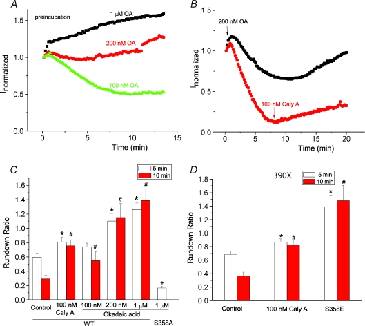
Figure 4. Dephosphorylation of S358 by PP2A regulates channel rundown.
A and B, time course of wild-type hBest1 currents recorded from cells pretreated with 100 nm (triangles), 200 nm (circles) and 1 μm (squares) okadaic acid (OA) for 2–5 min (A) and from cells treated with 200 nm OA (squares) or 100 nm calyculin A (circles) after establishing whole cell recording (B). C, effects of okadaic acid and calyculin A on channel rundown. Channel rundown was measured as the ratio of current amplitude 5 min (open columns) or 10 min (filled columns) after peak current to the peak amplitude (n= 4–12 cells; *P < 0.05 versus wild-type control with rundown for 5 min; #P < 0.05 versus wild-type control with rundown for 10 min; +P < 0.05 versus wild-type treated with 1 μm okadaic acid). D, effects of calyculin A and S358E mutation on channel rundown of 390X mutations. Channel rundown was measured as the ratio of current amplitude 5 min (open columns) or 10 min (filled columns) after peak current to the peak amplitude (n= 4–5 cells; *P < 0.05 versus control with rundown for 5 min; #P < 0.05 versus control with rundown for 10 min).

