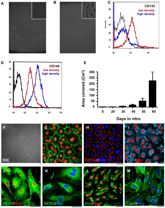
Figure 1. In vitro expansion, differentiation, and characterization of endothelial progenitor cells.
DIC images of endothelial progenitor cells (EPCs) during in vitro expansion (A) and differentiation (B). EPCs grow as highly proliferative and flattened cells during expansion (A) and differentiate into mature endothelial cells with cobble stone morphology as seen in panel B. FACS analysis of these cells during expansion (red color histogram) and after differentiation to mature endothelial cells (blue color histogram) is shown in (C,D). The black histograms in (C,D) represent the isotype controls. (E) demonstrates the equivalent surface area that could be endothelialized by EPCs obtained during in vitro expansion. Characterization of in vitro expanded and differentiated endothelial cells (F) shows that these cells are immunopositive for endothelial cell specific markers CD31, CD144, vWF,UEA1, and eNOS (G-K). Besides these endothelial markers they also produce Vimentin and caveolin1 (L and M) Bar = 10 µm.

