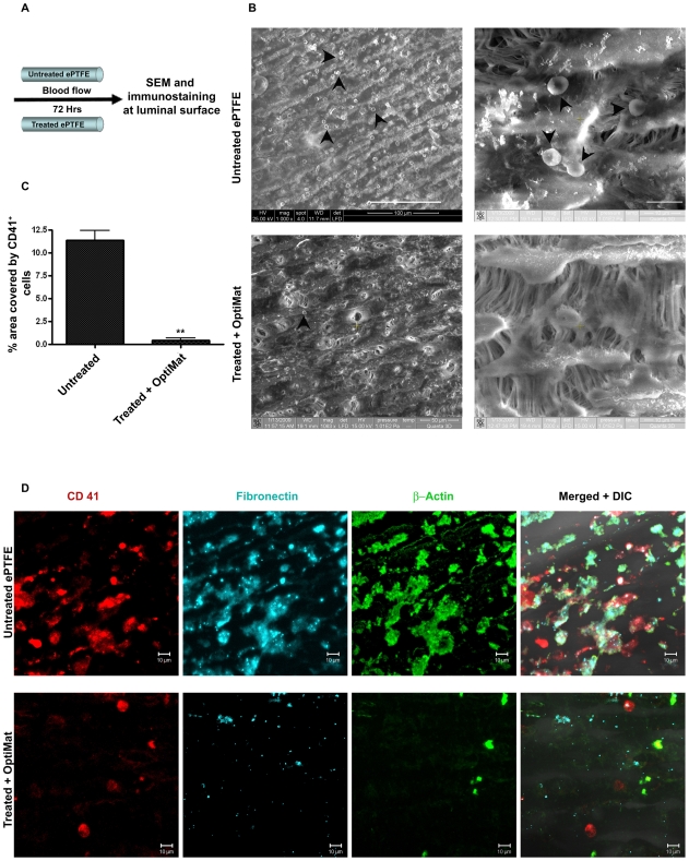
Figure 4. OptiMat coating to ePTFE grafts inhibit blood cells (platelets) adhesion.
ePTFE coated with OptiMat allow minimal platelets adhesion in blood flow condition in vitro. (A) Diagrammatic representation of blood flow through small diameter ePTFE tubings. After unidirectional blood flow scanning electron microscopy (SEM) (B) and immunostaining (D) were done to see blood platelets adhesion. OptiMat coated ePTFE tubes were observed with less number of blood cells (shown by arrow heads) adhered to them compared to untreated one (B). OptiMat coated tubes have less CD41 (platelets marker) +ve cells than untreated tubes (D). Percent area covered by CD41 +ve cells was calculated from randomly selected areas each of about 5000 µm2 of grafts (C). (D) also shows less fibronectin deposition (either from blood plasma or produced from platelets) and β-actin producing platelets on OptiMat coated tubes (D).

