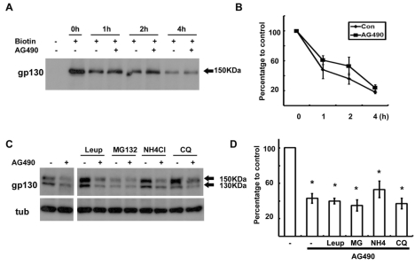
Fig. 3.
Degradation of gp130 was not enhanced by AG490. (A) Cell surface proteins were biotinylated at 4℃, and then cells were returned to 37℃ for the indicated time in the presence or absence of AG490 (50 µM). Protein extracts were precipitated with streptavidin-agarose beads, and analyzed by Western blotting with an antibody against gp130. (B) Quantitative results showing the change in biotinylated gp130 levels following AG490 treatment. (C) Cells were pretreated with protease inhibitors for 30 min and then cells were exposed to AG490 (50 µM) for 3 hrs. (D) Quantitative results showing the change in gp130 levels following AG490 treatment in the presence of several protease inhibitors. Leup; leupeptin, MG; MG132, NH4; NH4Cl, CQ; chloroquine. Data represent the means±S.E of three independent experiments. *p<0.05.

