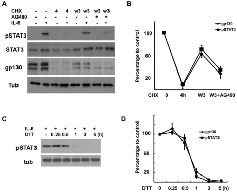
Fig. 6.
Correlation between gp130 levels and IL-6 response. (A) Cells were pretreated with CHX (10 µM) for 4 hrs, and then washed and reincubated in normal growth medium with or without AG490 (50 µM) for 3 hrs, and then IL-6 (50 ng/ml) was added for 15 min. The protein extracts were analyzed by Western blotting. (B) Quantitative results showing the changes in gp130 and pSTAT3 levels. Data represent the means±S.E of three independent experiments. (C) The cells were pretreated with dithiothreitol (DTT, 3 mM) for the indicated times and then stimulated with IL-6 for 15 min. Protein extracts were analyzed by Western blotting for pSTAT3. (D) Quantitative results showing the changes in gp130 and pSTAT3 levels in a time-dependent manner. Data represent the means±S.E of three independent experiments.

