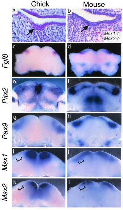
Figure 1.
Phenotypic and molecular comparison between putative chick dental lamina and mouse dental lamina. (a) Transverse section through stage 27 chick mandible showing a representative epithelial thickening (arrow), proposed to represent a vestigial dental lamina (16). Not all embryos examined exhibited such structures. (b) Transverse section of E14.5 Msx1-Msx2 double mutant molar tooth germ arrested at the lamina stage; the stage shown is developmentally equivalent to E11.5 in wild-type embryos. [Reproduced with permission from ref. 11. (Copyright 1998, Company of Biologists LTD).] (c–l) Molecular comparison between oral regions of stage 27 chick and E11.5 mouse mandibles. (c) Fgf8 expression in stage 27 chick mandibular epithelium. (d) Fgf8 expression in E11.5 mouse molar dental lamina. (e) Pitx2 expression in stage 27 chick mandibular epithelium. (f) Pitx2 expression in E11.5 mouse molar and incisor dental laminae. (g) Pax9 expression in stage 27 chick mandibular mesenchyme. (h) Pax9 expression in E11.5 mouse molar and incisor mesenchyme. (i–l) Differential Msx expression in chick and mouse mandibular mesenchyme. In chick, Msx1 (i) and Msx2 (k) expression is restricted to mesial mandibular mesenchyme and does not extend as far distally (bracketed). In mouse, Msx1 (j) and Msx2 (l) mesenchymal expression extends distally to the molar tooth-forming region (bracketed).

